

Oospores of P. agathidicida in the roots of kauri seedlings inoculated with P. agathidicida. The root has been cleared with potassium hydroxide and bleached with peroxide, before being stained with Trypan Blue
Photo Gallery
Site will be retired 9/1/2026
This site is no longer being developed and will be retired on September 1, 2026. Please contact us if you have any questions or would like to provide support to continue the project.
|
P. agathidicida oospores 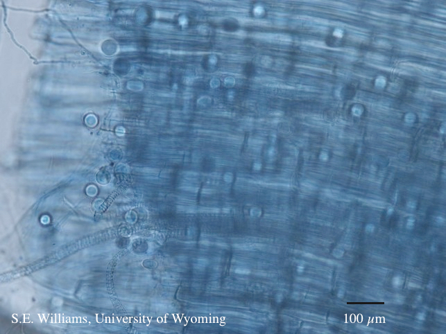 |
P. pluvialis symptoms 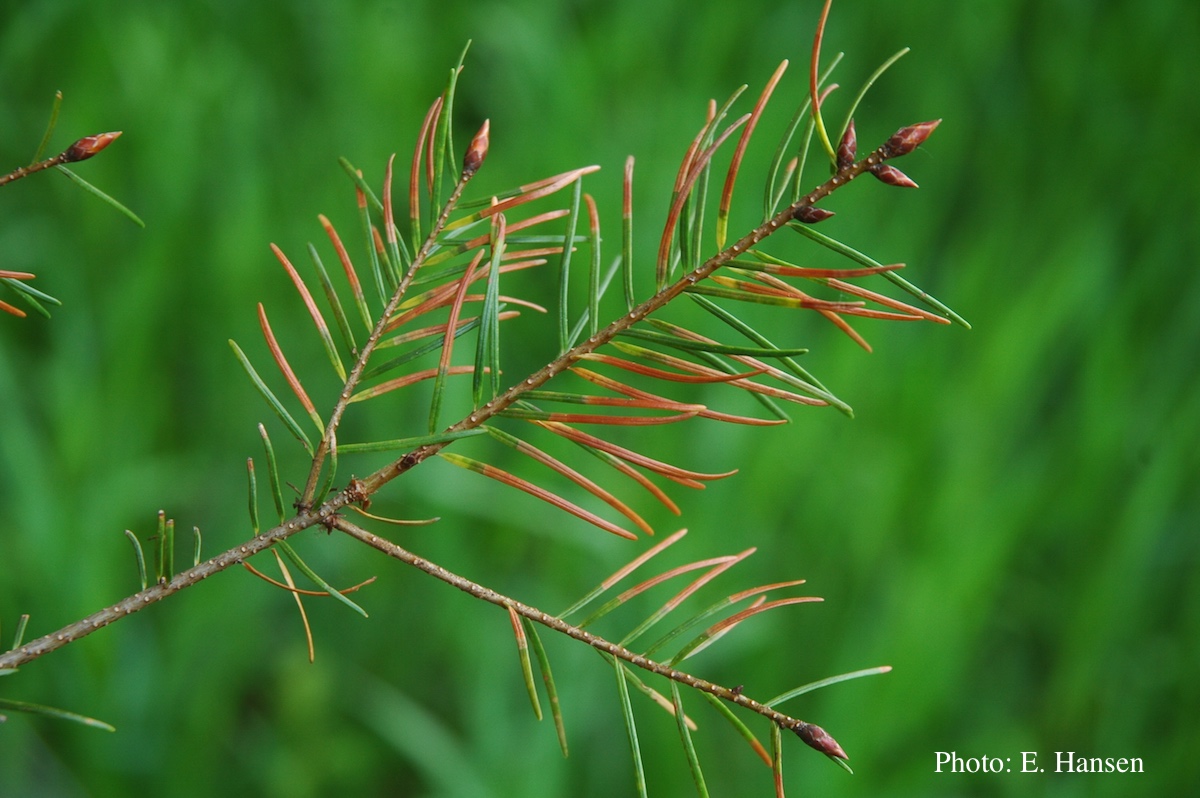 Symptoms of red needle cast on Douglas-fir needles |
P. austrocedrae - hyphal swellings 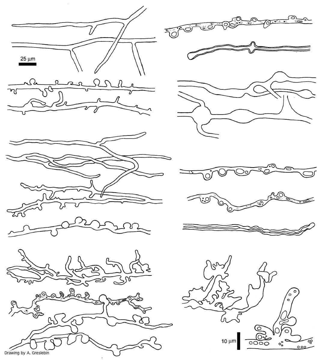 Morphology of hyphae of Phytophthora austrocedrae, from Greslebin et al. 2007 |
|
P. cambivora sporangium with internal extended proliferation 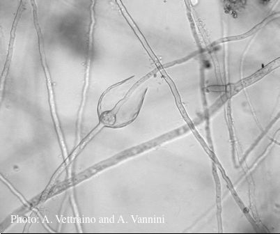 Empty sporangia showing internal extended proliferation |
P. nicotianae symptoms 2 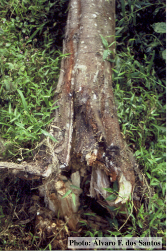 Symptoms of gummosis on black wattle (Fitopatol. bras. 2005) |
P. siskiyouensis canker on Italian alder 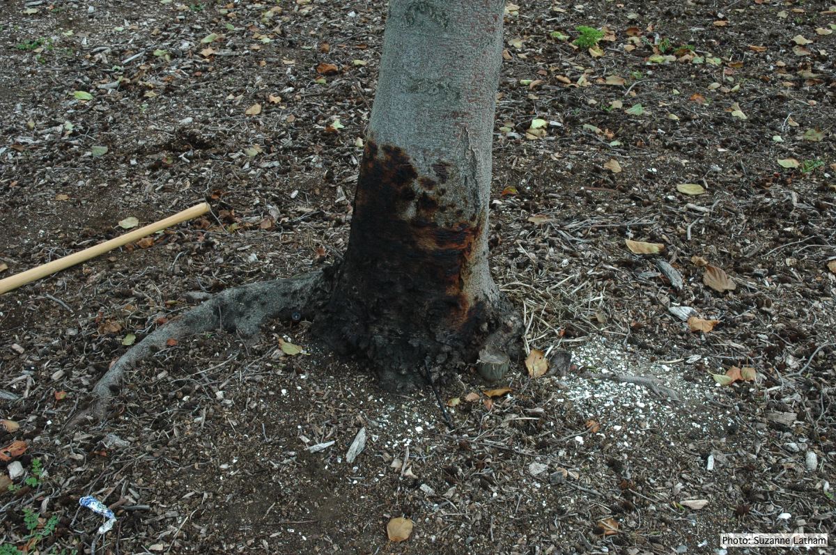 Bleeding canker at the base of a tree and a sprinkler emitter (arrow) adjacent to the trunk |
|
P. palmivora sporangia, chlamydospores, hyphae 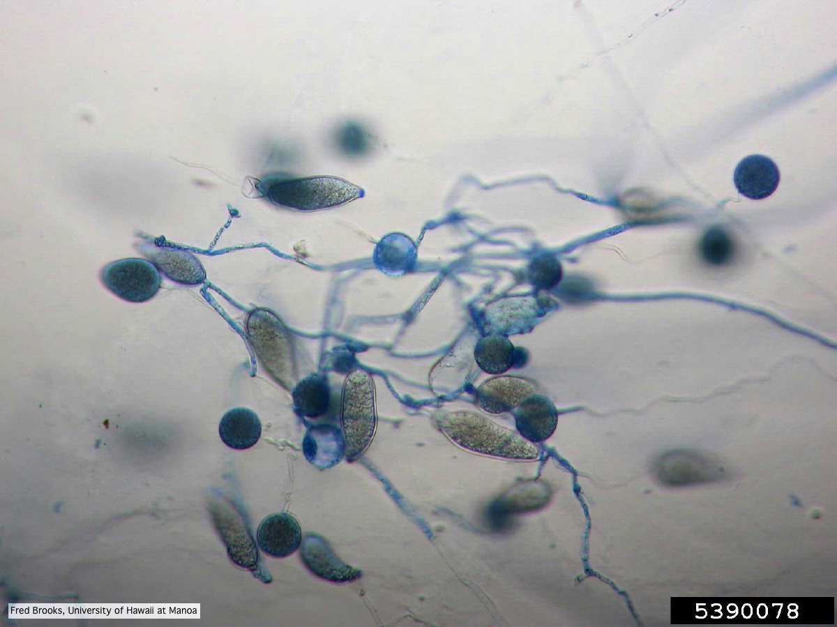 Sporangia (sporangiospores), chlamydospores, and hyphae stained with Cotton Blue |
P. siskiyouensis canker on Italian alder 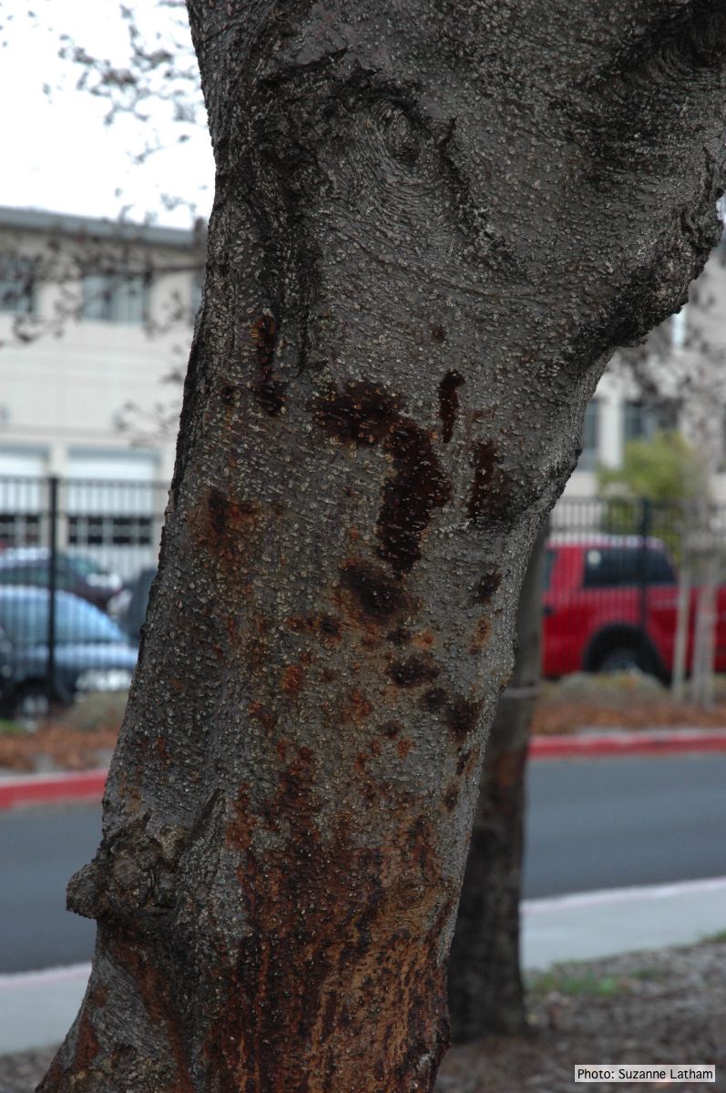 Phytophthora collar rot on Italian alder trees: an isolated bleeding canker on the trunk |
Dying Port Orford Cedar trees 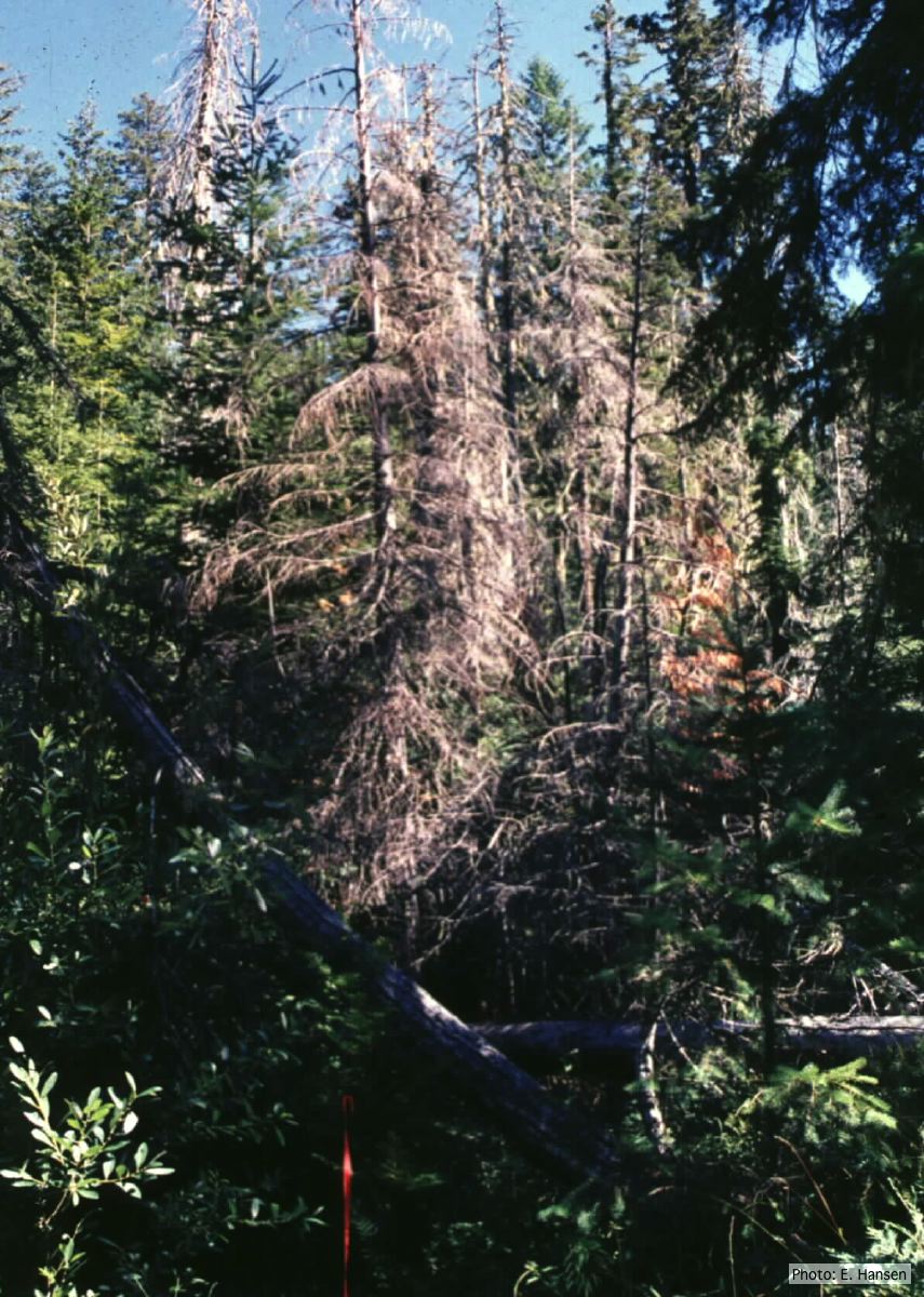 Dead Chamaecyparis lawsoniana trees |
|
P. kernoviae colony morphology 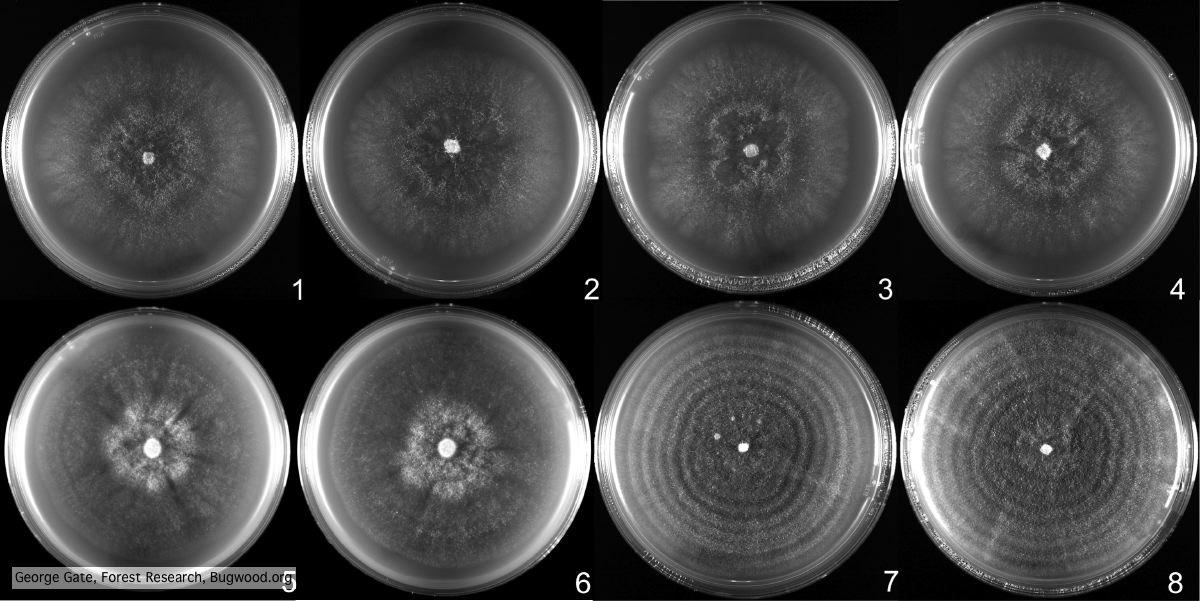 From Mycol.Res 109, 853-859; growth on CA under different conditions |
P. tentaculata microscopic characteristics 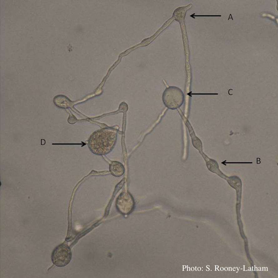 Hyphal swellings occuring at branching points of Mycelium (A), Intercalary hyphal swellings (B), Chlamydospore (C ), Sporangia (D) |
P. pluvialis hyphal swellings 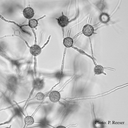 P. pluvialis hyphal swellings in water |