

Globose chlamydospore of P. boehmeriae
Photo Gallery
Site will be retired 9/1/2026
This site is no longer being developed and will be retired on September 1, 2026. Please contact us if you have any questions or would like to provide support to continue the project.
|
P. boehmeriae chlamydospore 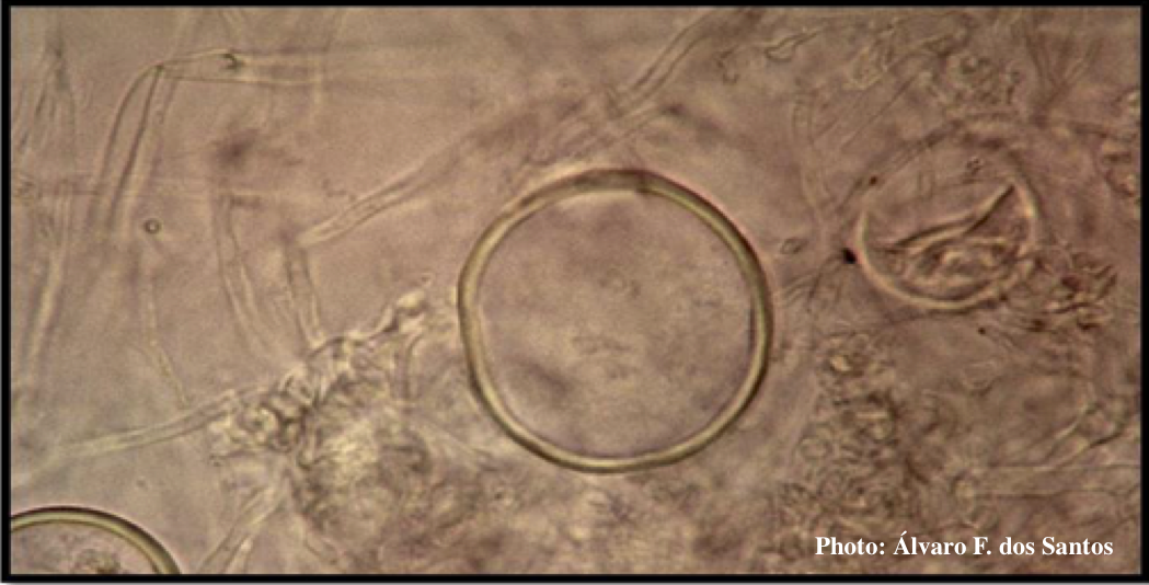 |
P. cactorum sporangia 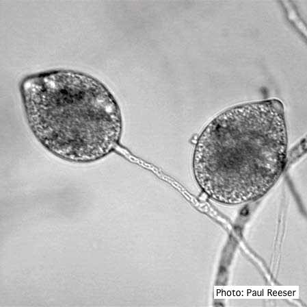 Broadly ovoid, papillate sporangia in water. |
P. cinnamomi on Fraser fir 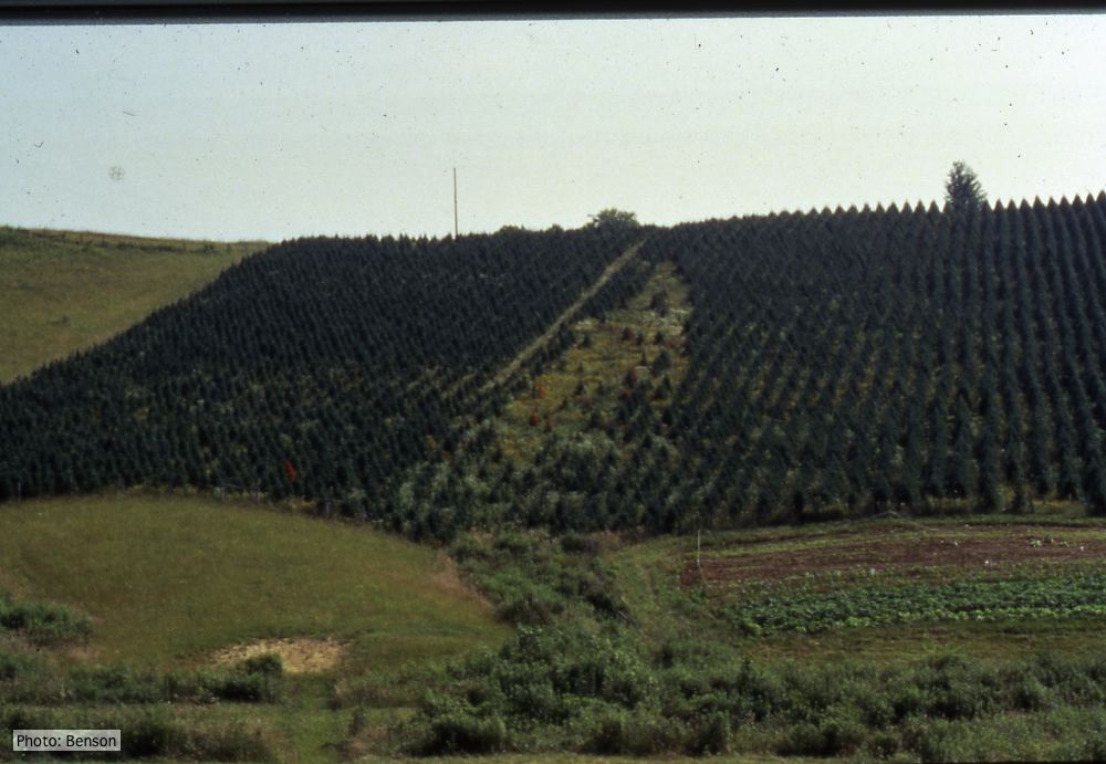 Frasier fir Christmas trees, North Carolina |
|
P. cambivora on dead and dying chinquapin 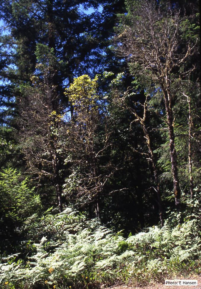 Dead and dying chinquapin infected with P. cambivora |
P. palmivora sporangia, chlamydospores, hyphae 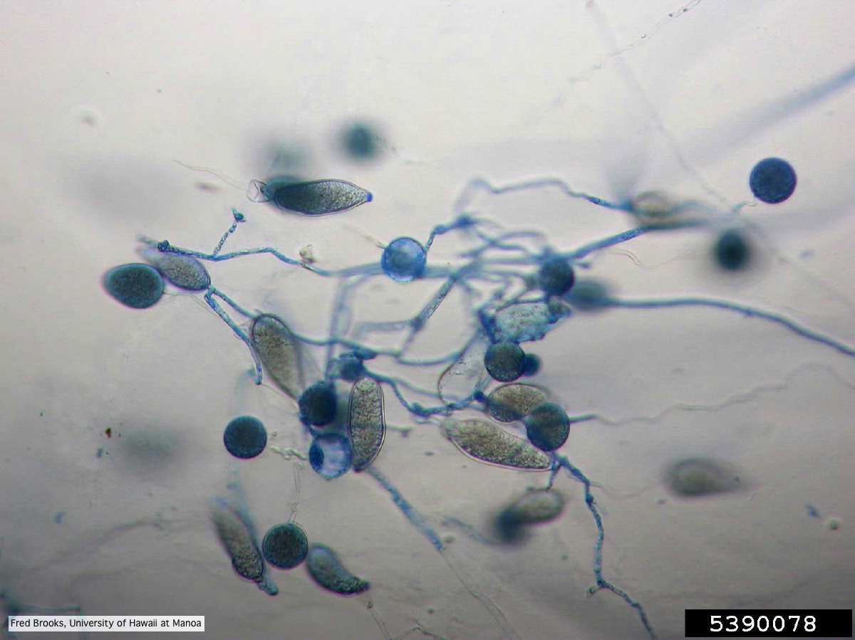 Sporangia (sporangiospores), chlamydospores, and hyphae stained with Cotton Blue |
Comparative gametangial morphology of Phytophthora Clade 5 species 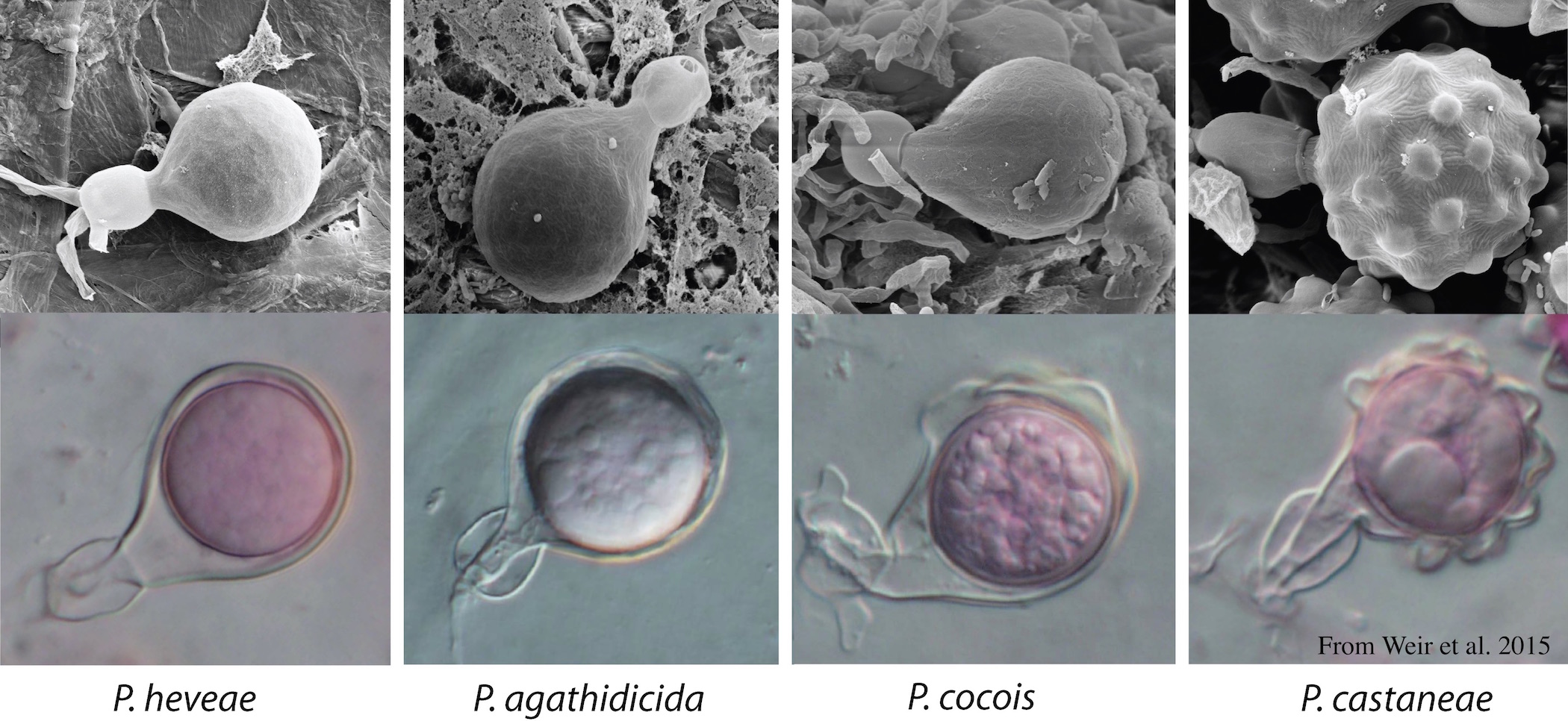 Comparative gametangial morphology of Phytophthora Clade 5 species, with SEM (top) and light microscopy (bottom). P. heveae has smooth walled oogonia with funnel-shaped, amphigynous antheridia. P. agathidicida has mildly stipulate oogonia with globose amphigynous antheridia. P.cocois has mildly bullate oogonia with reflexed amphigynous antheridia. P. castaneae has coarsely bullate oogonium with rugose protuberances and narrow amphigynous antheridia (Weir et al. 2015). |
|
P. pinifolia on Pinus radiata 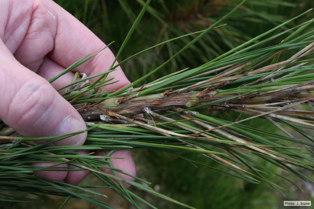 Pinus radiata needles, note “black line” symptom near needle bases |
Mal del ciprés, dead and dying trees along river 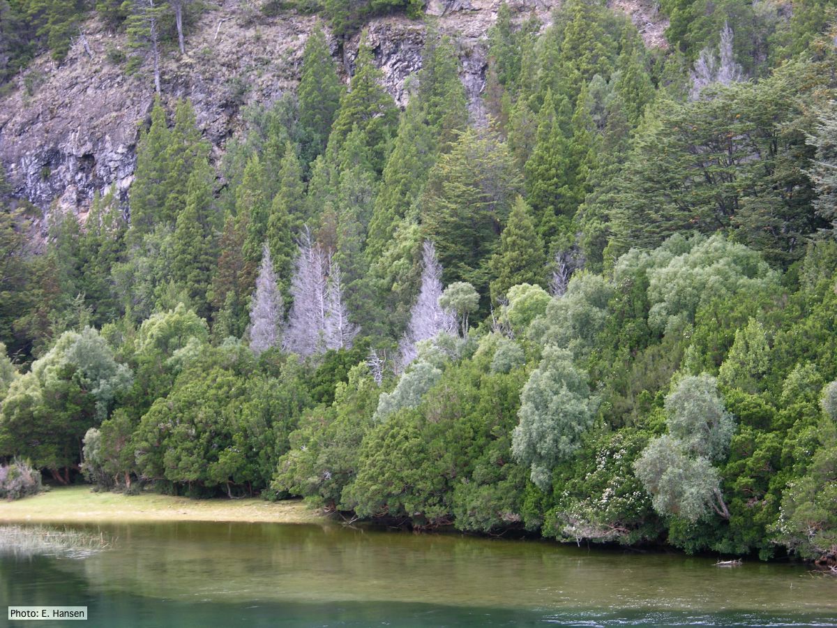 Mal del ciprés, dead and dying trees along river |
Necrotic lesion in phloem caused by P. austrocedrae 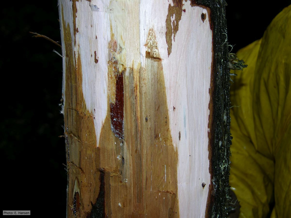 Necrotic lesion in phloem with resin pocket caused by P. austrocedrae |
|
P. cambivora oogonium 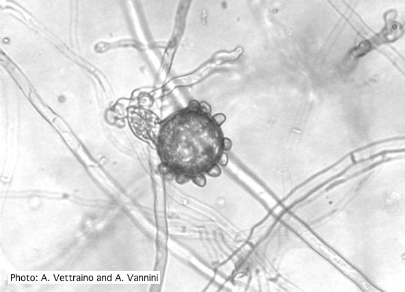 Bullate oogonium with amphyginous antheridium |
P. pinifolia sporangia 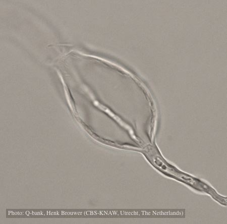 Sporangium with internal proliferation, photo from Q-bank, used with permission. |
Growth of P. arenaria on CA 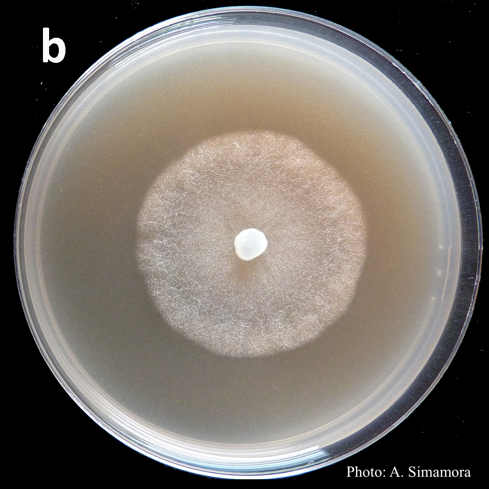 Colony morphology of Phytophthora arenaria after 7 days at 20°C on carrot agar |