

Jogger running over a plastic-reinforced, foam-mat containing a 2% solution of Trigene™ Advance (quaternary ammonium compound) as part of a cross-country event, in the Waitakere Regional Park
Photo Gallery
Site will be retired 9/1/2026
This site is no longer being developed and will be retired on September 1, 2026. Please contact us if you have any questions or would like to provide support to continue the project.
|
Mat to control spread of P. agathidicida 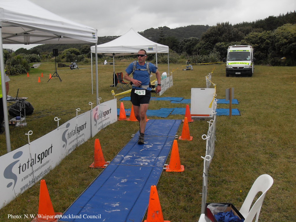 |
P. cambivora disease symptoms 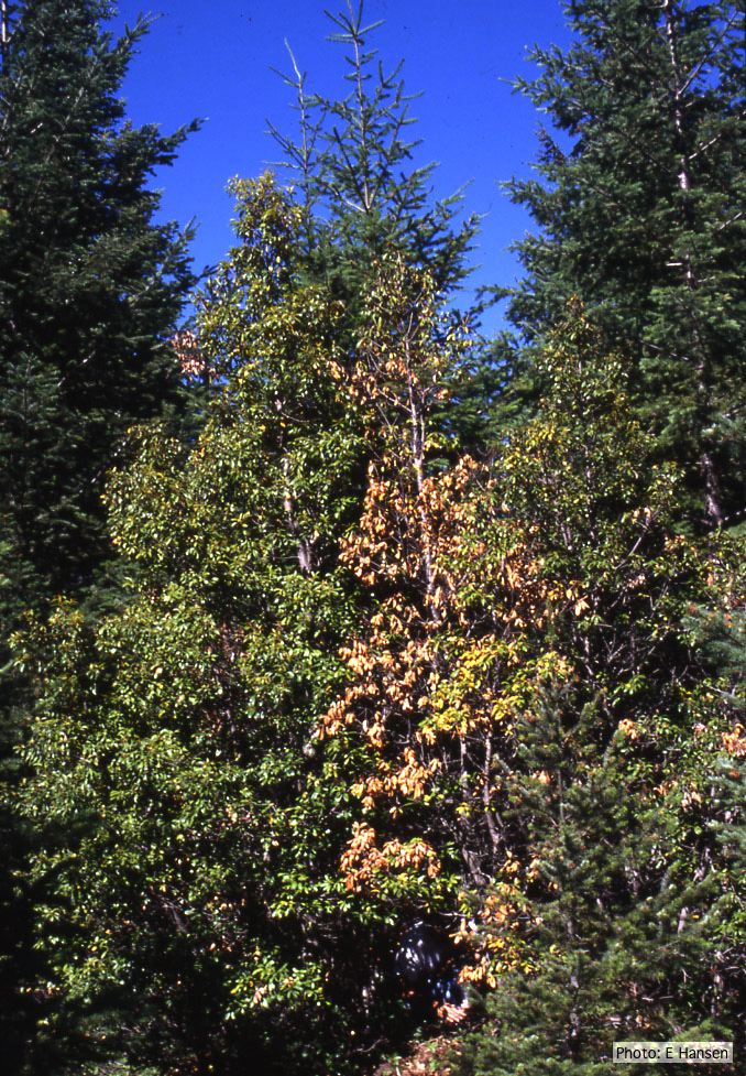 Dead and dying chinquapin infected with P. cambivora |
P. pinifolia on Pinus radiata 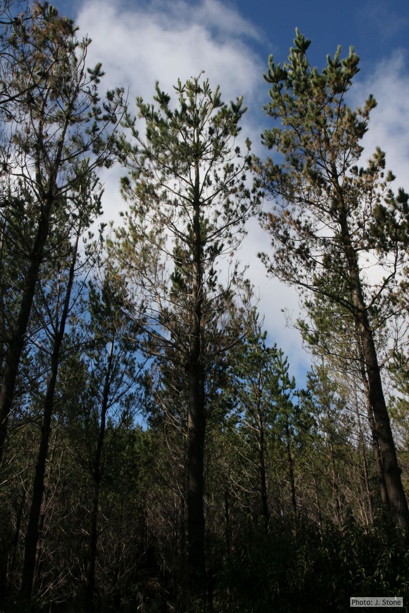 Pinus radiata impacted by DFP, note healthy new growth |
|
P. arenaria disease symptoms on Banksia landscape 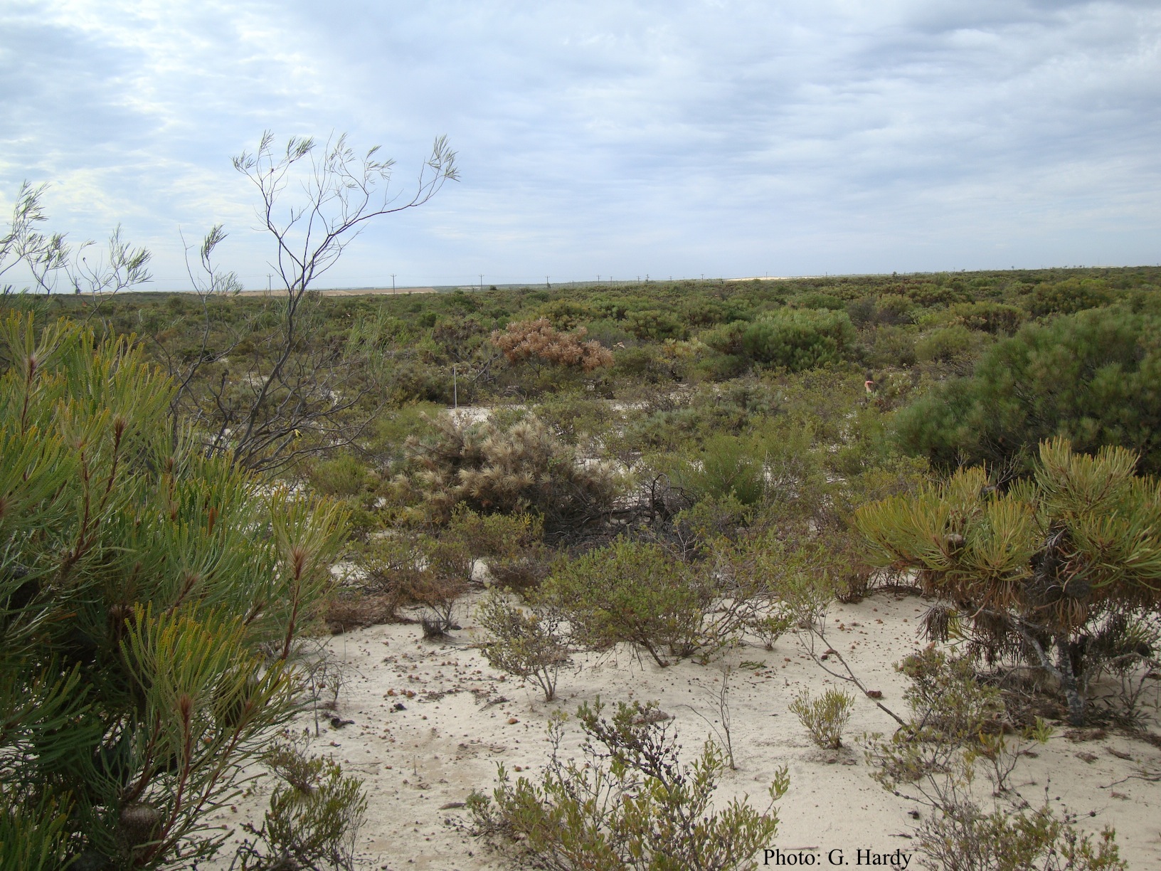 Dead Banksia sp. in a Kwongan heathland on mineral sand near Eneabba, Western Australia recently killed by root and collar rot caused by Phytophthora arenaria |
P. pluvialis symptoms on Douglas-fir needles 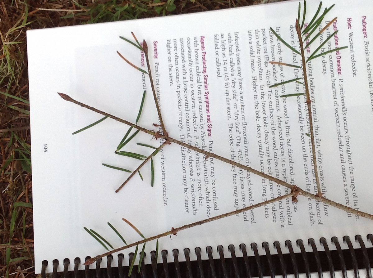 Symptoms of red needle cast on Douglas-fir needles |
P. nicotianae symptoms 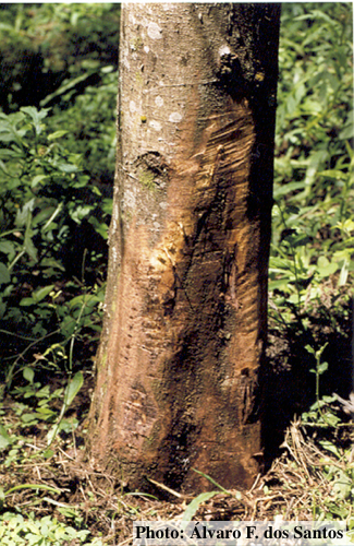 Symptoms of gummosis on black wattle (Fitopatol. bras. 2005) |
|
P. megasperma oogonium 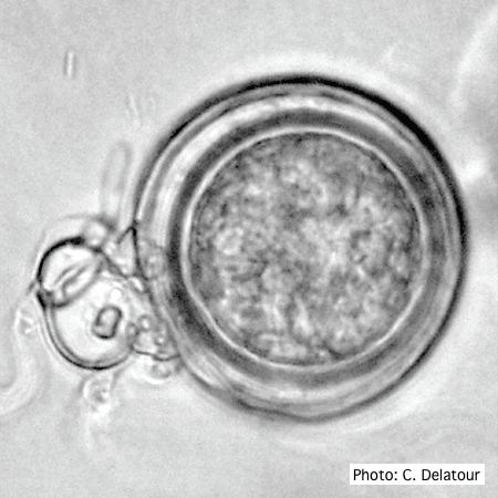 Oogonium with paragynous antheridia applied close to the ogonial stalk. |
P. pinifolia on Pinus radiata 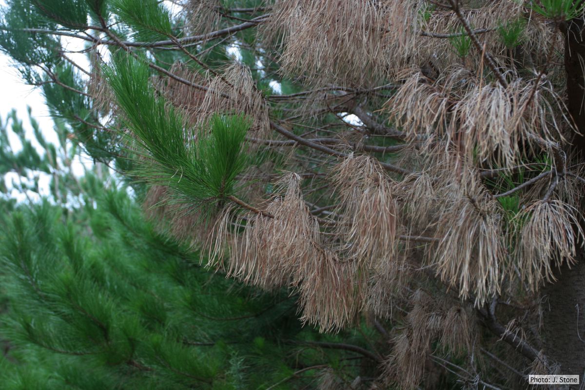 Pinus radiata dead needles caused by DFP with healthy new growth from DFP |
P. ramorum leaf symptoms on tan oak 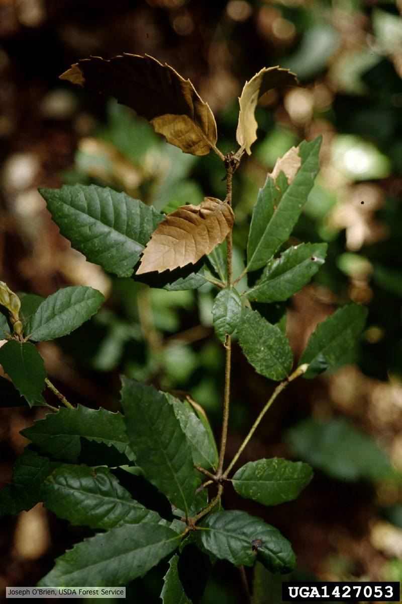 Tip symptoms on tanoak seedling (Notholithocarpus densiflorus). |
|
P. pseudotsugae colony morphology on PDA 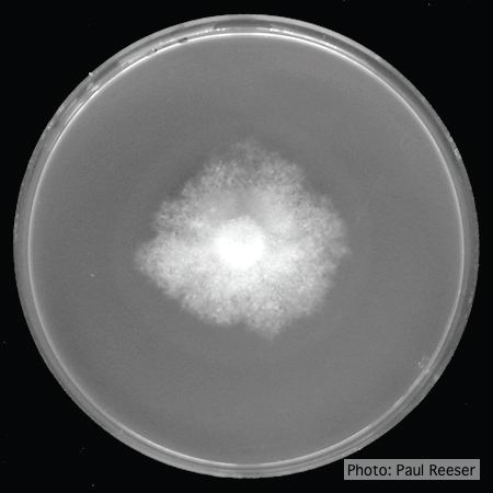 P. pseudotsugae colony growth on PDA agar |
Comparative gametangial morphology of Phytophthora Clade 5 species 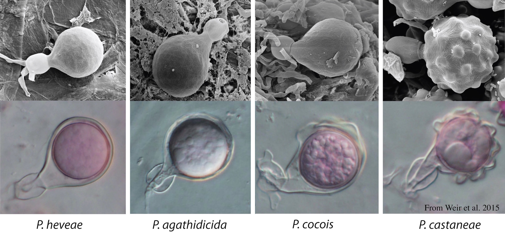 Comparative gametangial morphology of Phytophthora Clade 5 species, with SEM (top) and light microscopy (bottom). P. heveae has smooth walled oogonia with funnel-shaped, amphigynous antheridia. P. agathidicida has mildly stipulate oogonia with globose amphigynous antheridia. P.cocois has mildly bullate oogonia with reflexed amphigynous antheridia. P. castaneae has coarsely bullate oogonium with rugose protuberances and narrow amphigynous antheridia (Weir et al. 2015). |
P. pluvialis on Pinus radiata in New Zealand 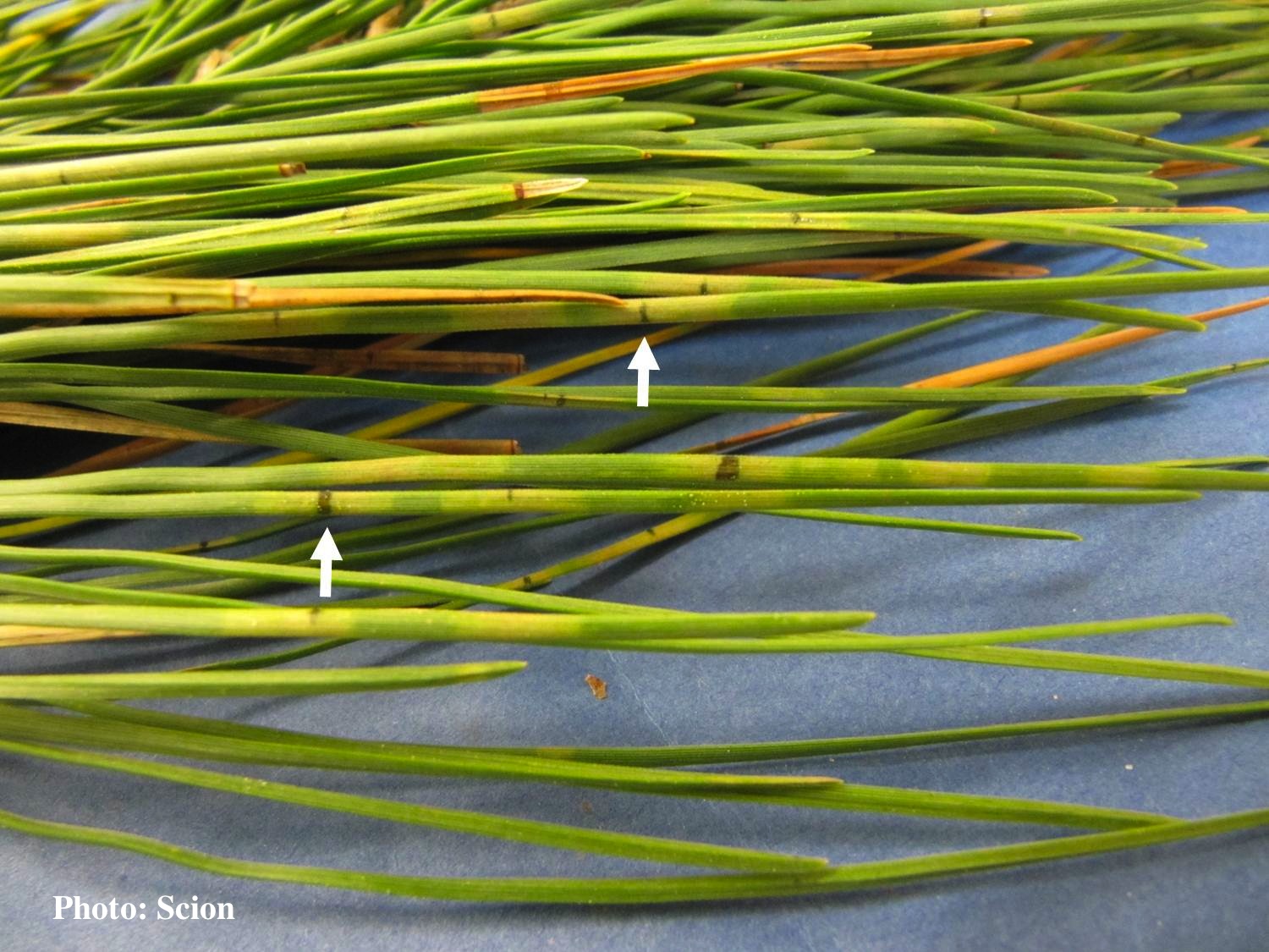 Pinus radiata needles showing colour changes following infection with red needle cast disease. The tissues around the initial infection at the base or along the needle senesce, and change yellow and then brown as indicated by the arrows before the needles cast. |