
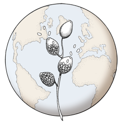

Colony morphology on PDA at 14 days
Photo Gallery
Site will be retired 9/1/2026
This site is no longer being developed and will be retired on September 1, 2026. Please contact us if you have any questions or would like to provide support to continue the project.
|
P. cambivora colony morphology on PDA 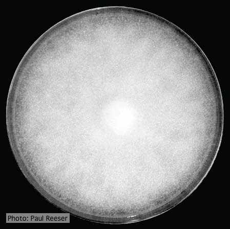 |
P. nicotianae sporangia 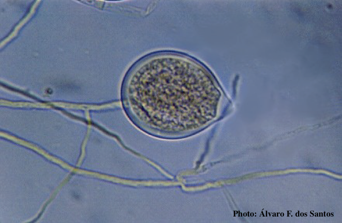 Noncaducous sporangium showing ovoid shape and papillate condition. (Fitopatol. bras. 2005) |
P. agathidicida lesion on kauri tree 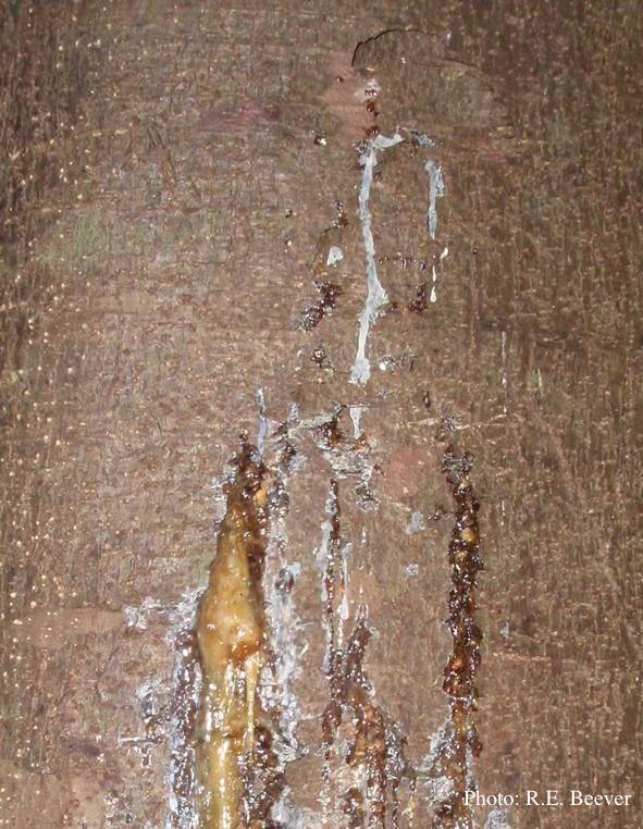 Gum oozing out of longitudinal lesion |
|
P. cambivora symptoms 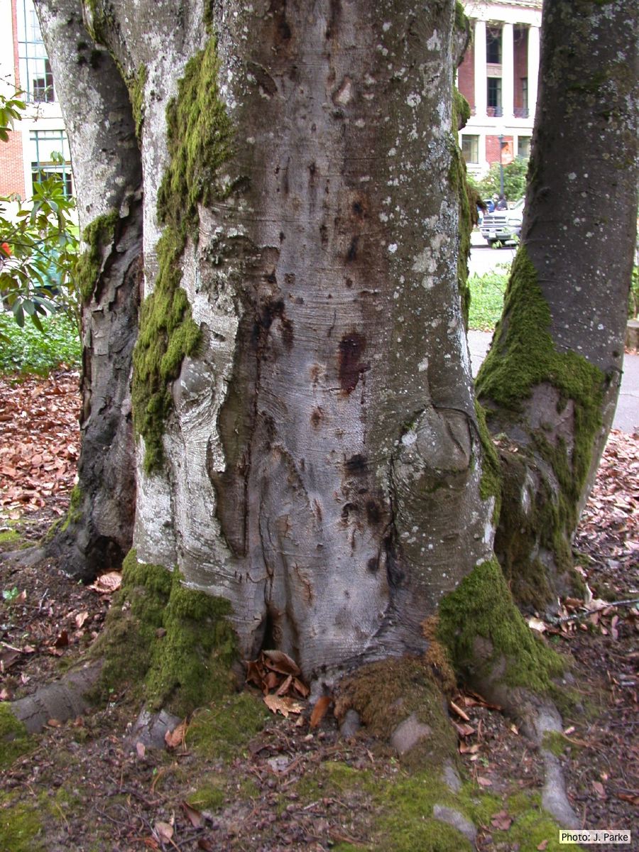 P. cambivora on Fagus sylvatica bole |
Growth of P. palmivora on CMA 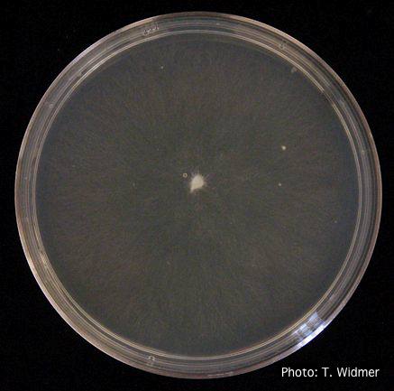 Growth of P. palmivora on corn meal agar |
P. cactorum bleeding canker 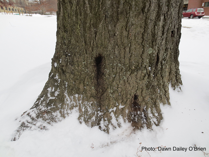 Bleeding canker on red oak (Quercus rubra) |
|
P. ramorum colony morphology on CMA PARP 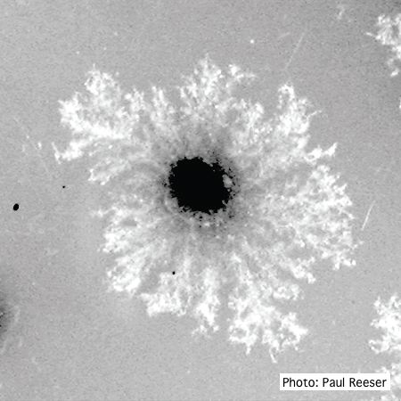 P. ramorum colony morphology on CMA PARP |
P. pluvialis symptoms on Douglas-fir 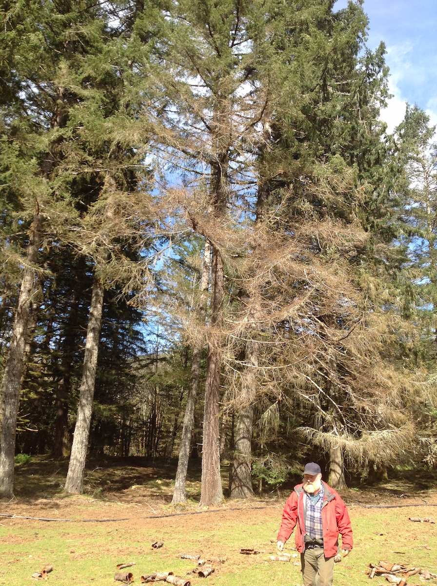 Red needle cast symptoms on Douglas-fir in western Oregon, 2015 |
P. palmivora symptoms on fruit 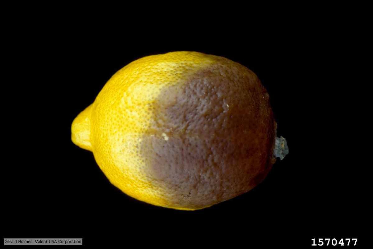 Brown rot on a lemon fruit caused by Phytophthora palmivora. |
|
P. pinifolia sporangium 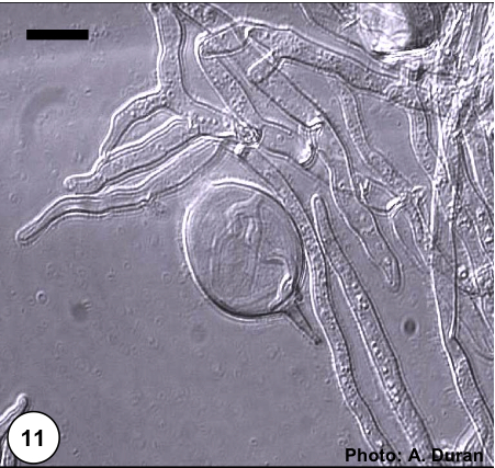 Non- papillate and caducous sporangia of Phytophthora pinifolia isolated from the infected P. radiata needles. |
P. ramorum colony morphology on PDA 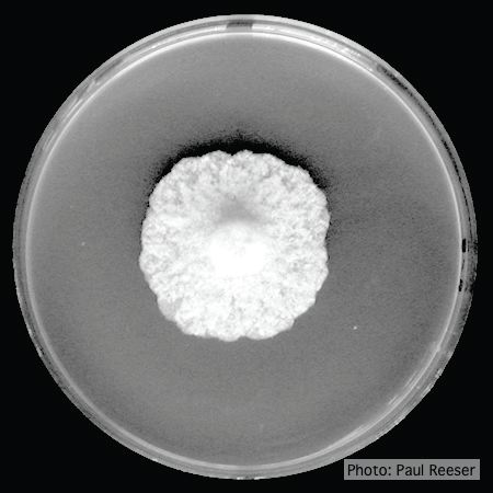 P. ramorum colony morphology on PDA |
P. katsurae sporangia 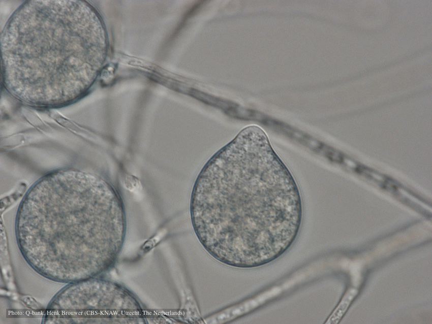 Papillate, non-caducous sporangia; photo used with permission from Q-bank |