

Close-up of margin area of bole lesions under the bark of a bleeding canker
Photo Gallery
Site will be retired 9/1/2026
This site is no longer being developed and will be retired on September 1, 2026. Please contact us if you have any questions or would like to provide support to continue the project.
|
P. siskiyouensis bleeding canker 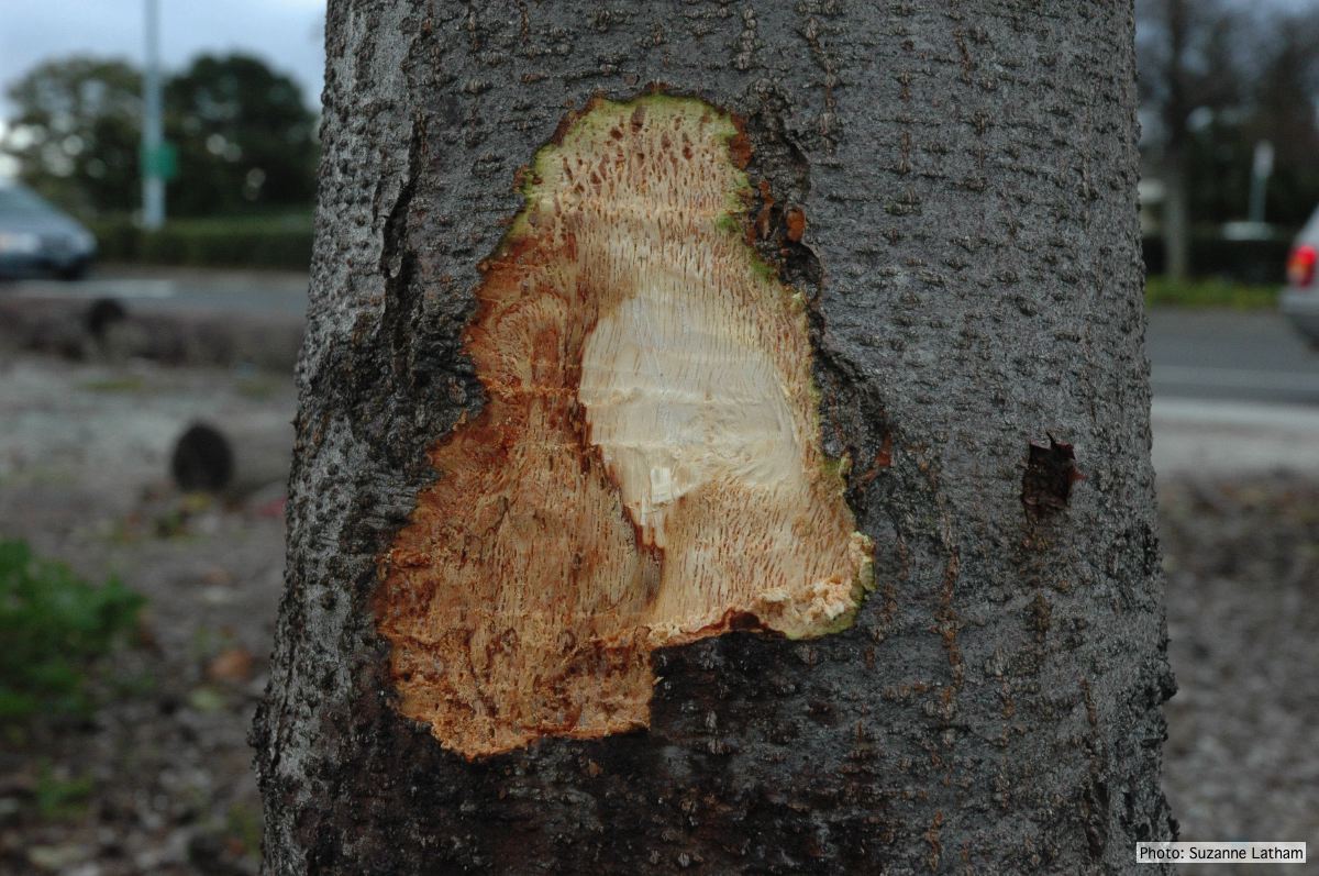 |
P. cambivora on dead and dying chinquapin 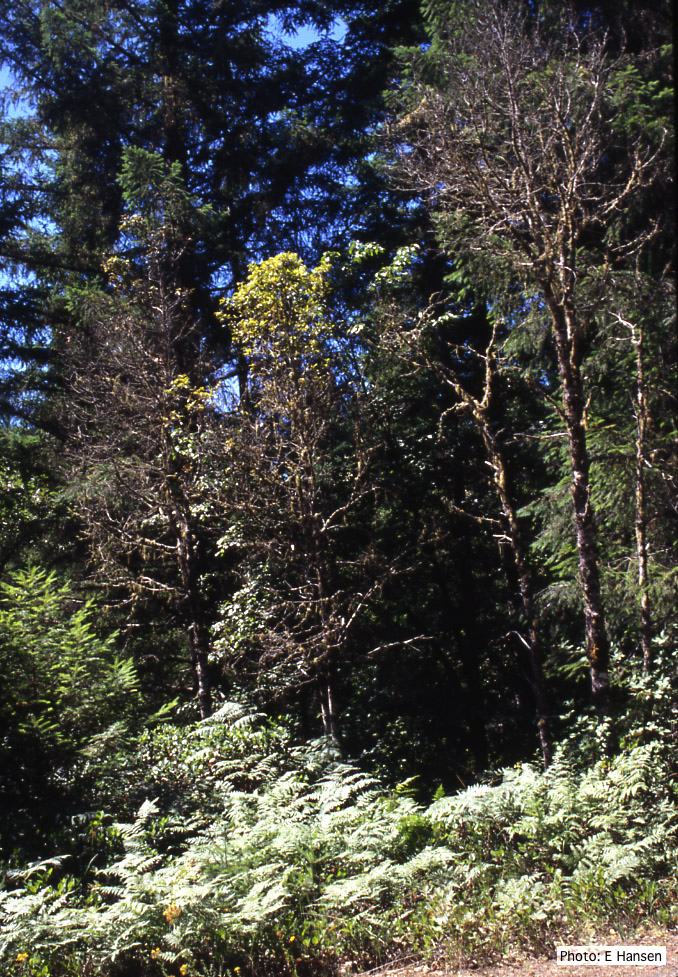 Dead and dying chinquapin infected with P. cambivora |
P. pseudosyringae oogonium 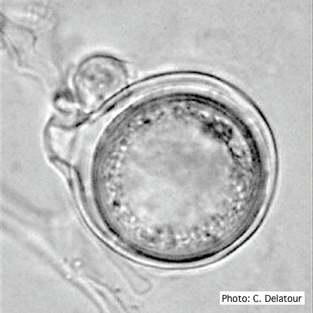 Oogonium with paragynous antheridium in agar |
|
P. pseudosyringae colony morphology on V8 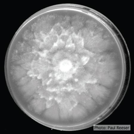 Colony morphology on V8 at 14 days |
P. lateralis on Port Orford cedar 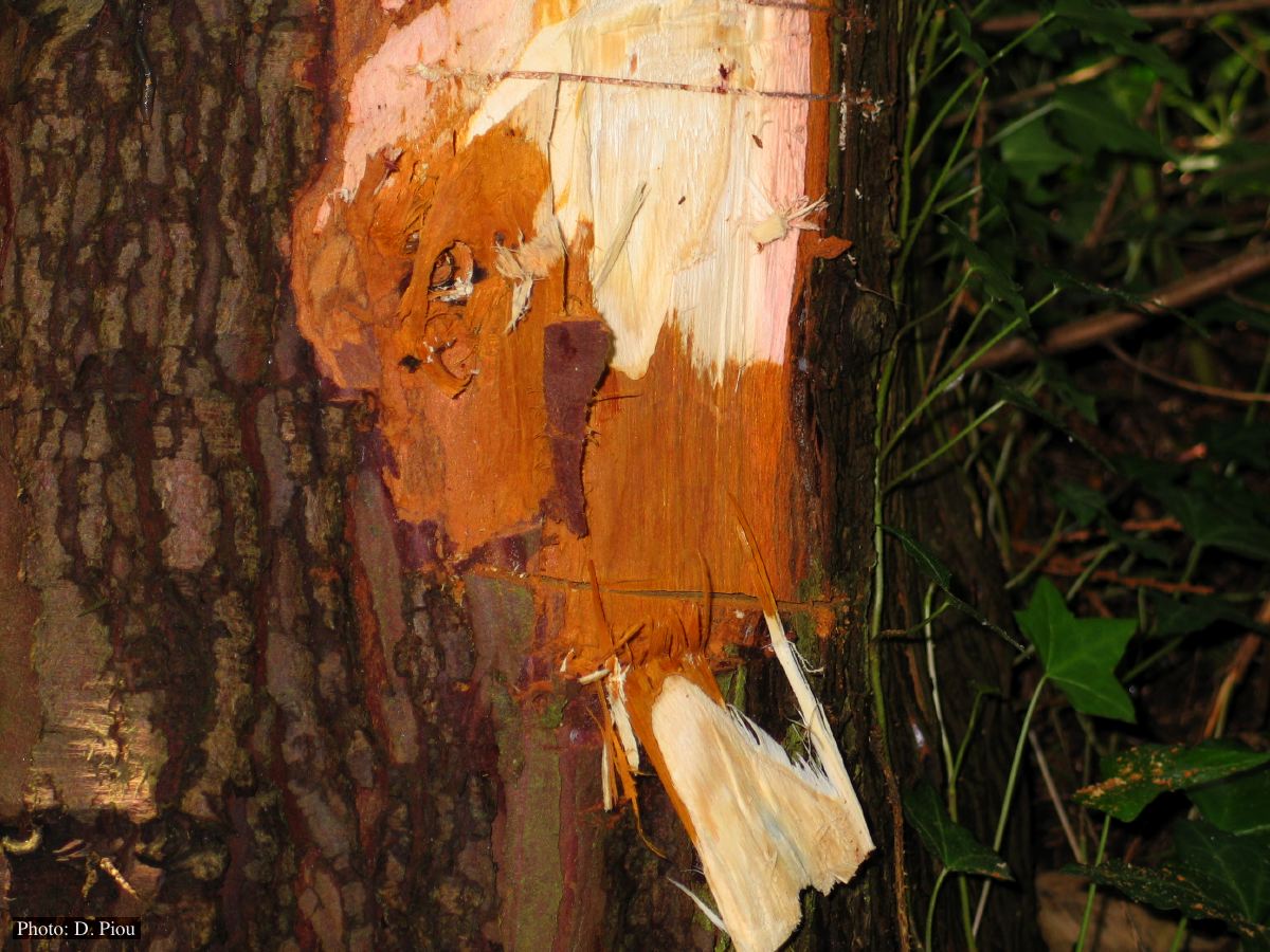 Collar lesion on Chaemacyparis lawsoniana in Landrévarzec, France |
P. pseudotsugae colony morphology on V8 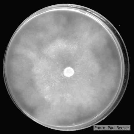 P. pseudotsugae colony growth on V8 agar |
|
P. lateralis on Port Orford cedar 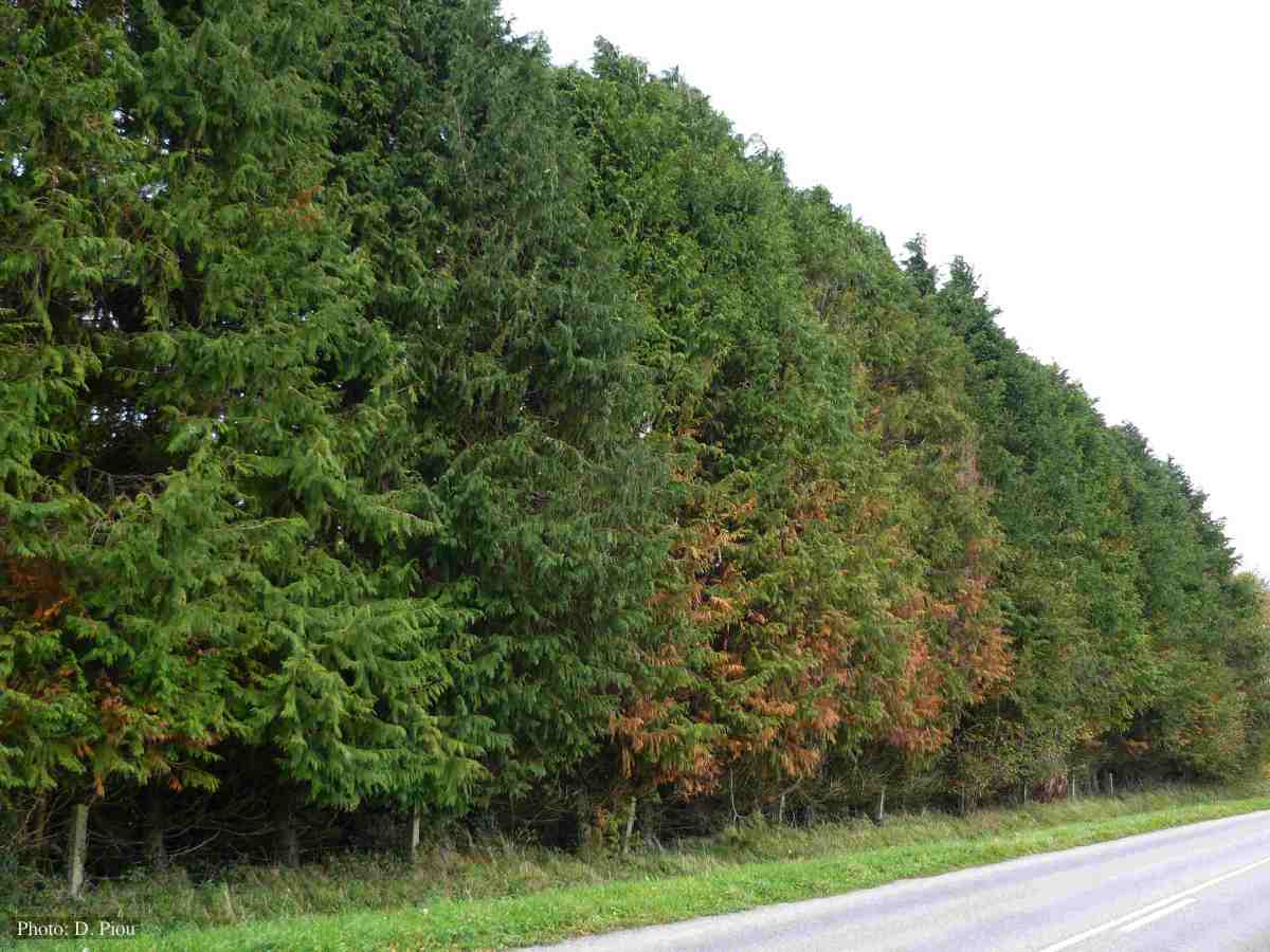 Atypical decline caused by aerial infections in Scaër, France |
P. cryptogea sporangia 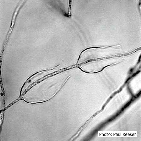 Sporangiophore showing internal proliferation through empty sporangia after zoospore release |
P. tentaculata disease symptoms on California mugwort 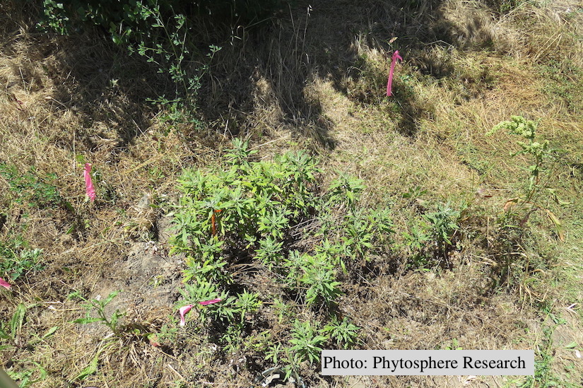 Outplanted California mugwort (Artemisia douglasiana) infected with P. tentaculata, 4.5 years after planting. Plant shows stunting and chlorosis. (P. cryptogea and P. lacustris were also baited from roots/soil of this plant). |
|
P. pluvialis symptoms on Douglas-fir 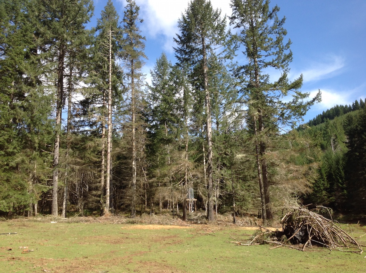 P. pluvialis symptoms of red needle cast on Douglas-fir, western Oregon 2015 |
P. alni symptoms on European Alder 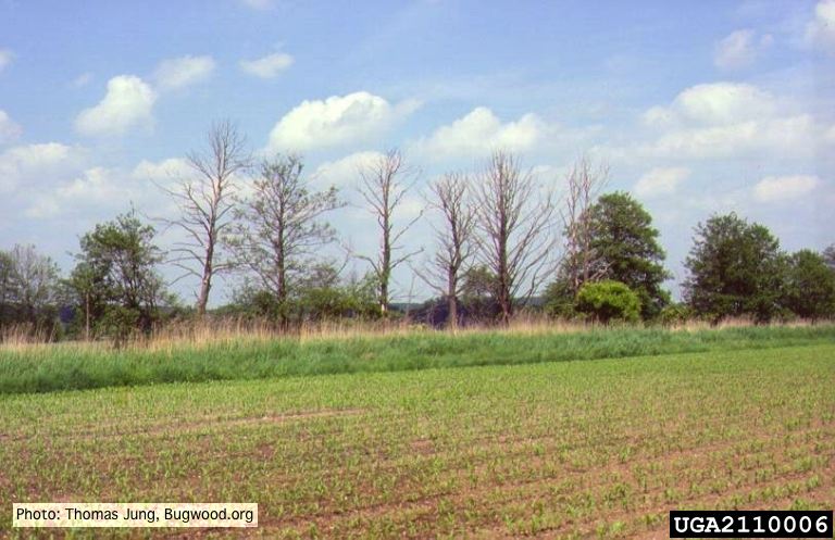 Mature, riparian common alder (A. glutinosa) stand with high impact of Phytophthora root and collar rot. |
P. alni oogonia subspecies and variants 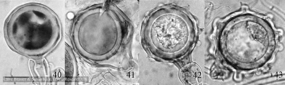 Fig. 40. P. alni subsp. uniformis. Fig. 41. P. alni subsp. multiformis German variant. Fig. 42. P. alni subsp. alni. Fig. 43. |