

Colony morphology on PDA at 7 days
Photo Gallery
Site will be retired 9/1/2026
This site is no longer being developed and will be retired on September 1, 2026. Please contact us if you have any questions or would like to provide support to continue the project.
|
P. megasperma colony morphology on PDA 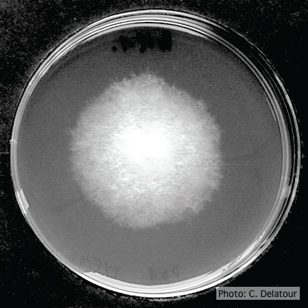 |
P. pseudosyringae colony morphology on V8 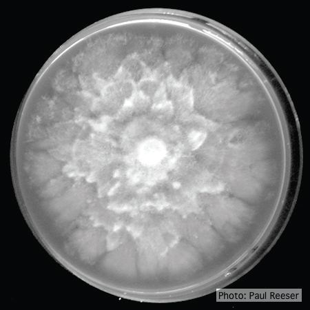 Colony morphology on V8 at 14 days |
P. siskiyouensis oogonium with amphigynous antheridium 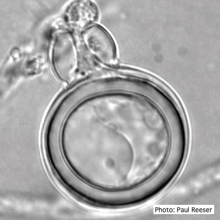 P. siskiyouensis oogonium with amphigynous antheridium |
|
P. lateralis on Port Orford cedar 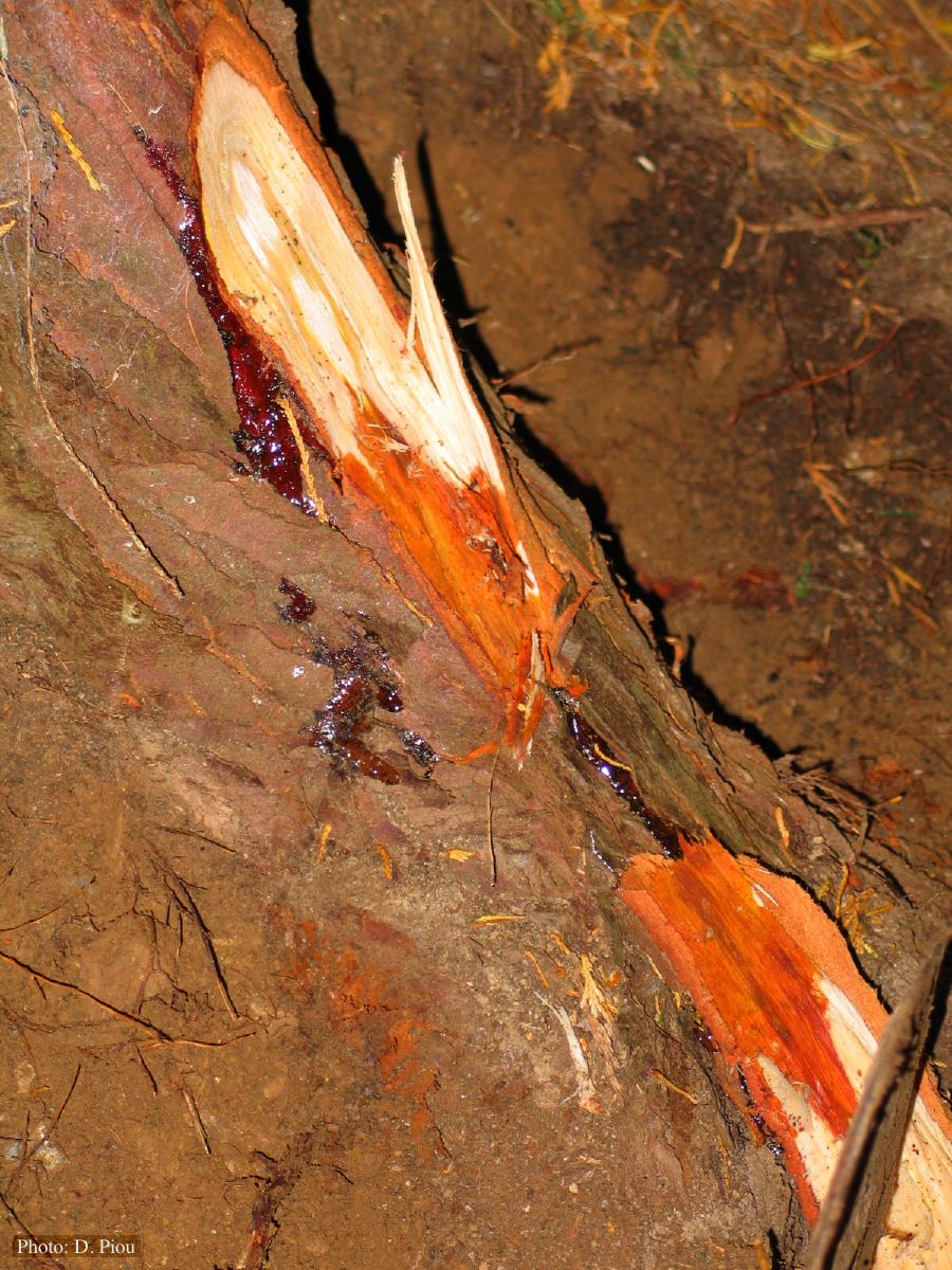 Small root lesions on Chaemacyparis lawsoniana |
P. palmivora symptoms on fruit 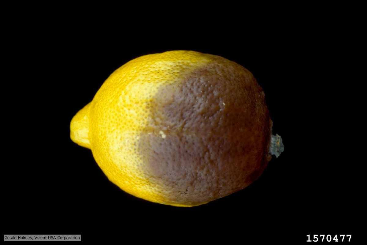 Brown rot on a lemon fruit caused by Phytophthora palmivora. |
P. pinifolia on Pinus radiata 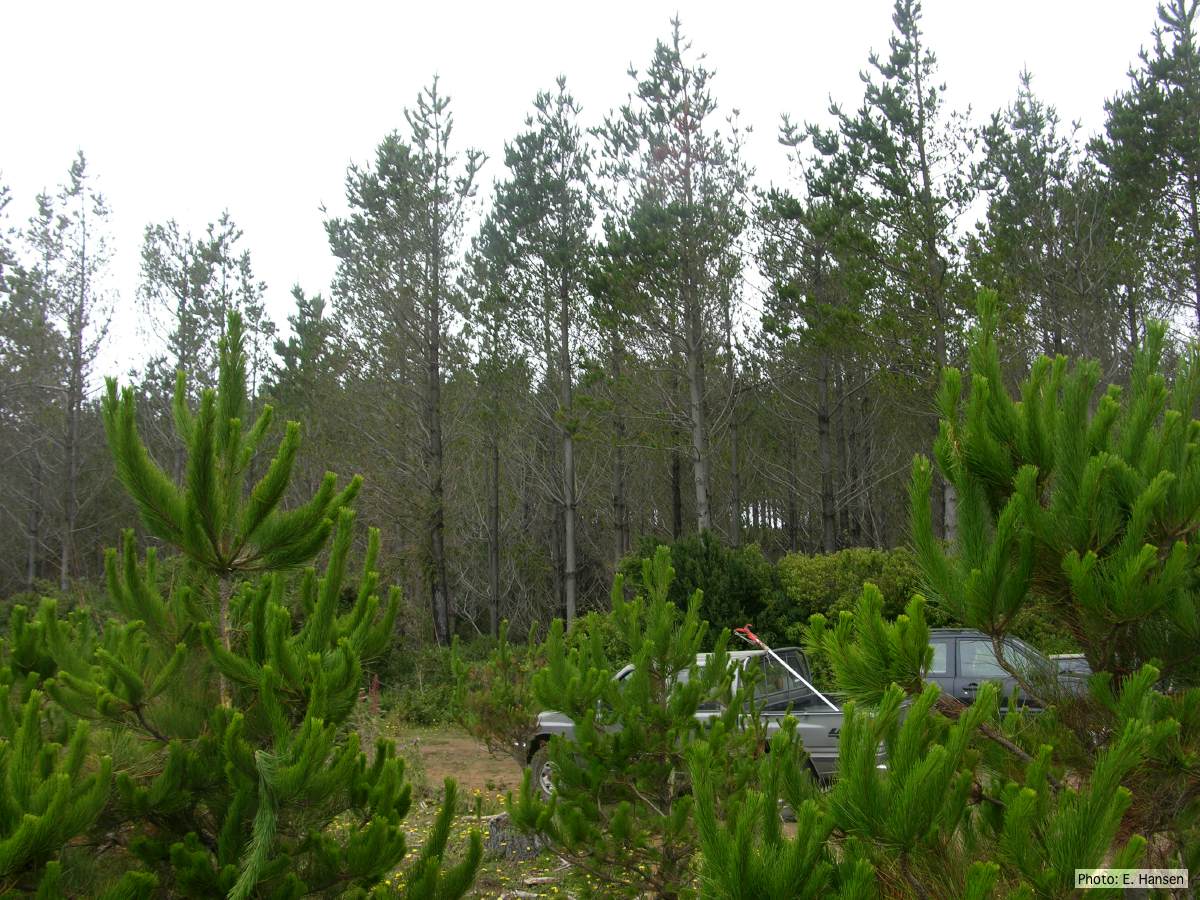 Pinus radiata stand, note Defoliation and regrowth |
|
P. pseudotsugae sporangium 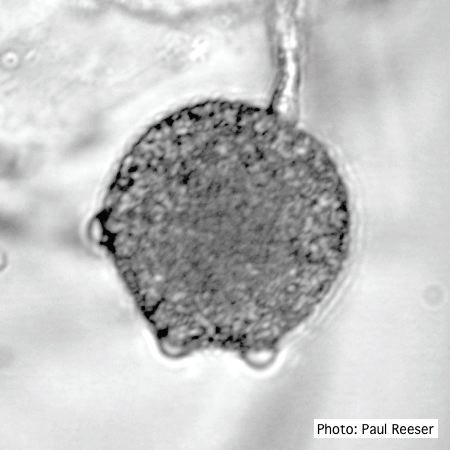 Broadly ovoid, papillate sporangium in water |
P. megasperma colony morphology on V8 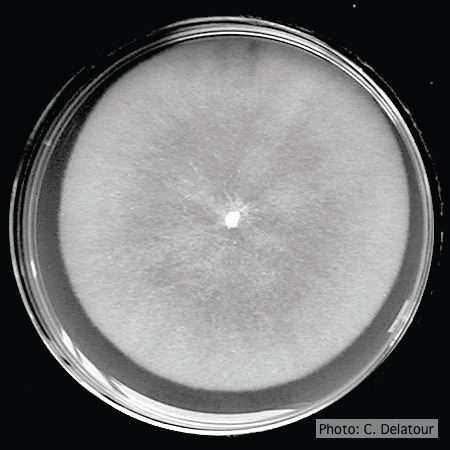 Colony morphology on V8 at 7 days |
P. cambivora oogonium 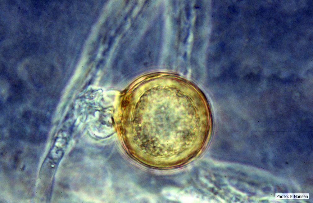 P. cambivora oogonium with antheridium |
|
Phytophthora chlamydospora sporangium 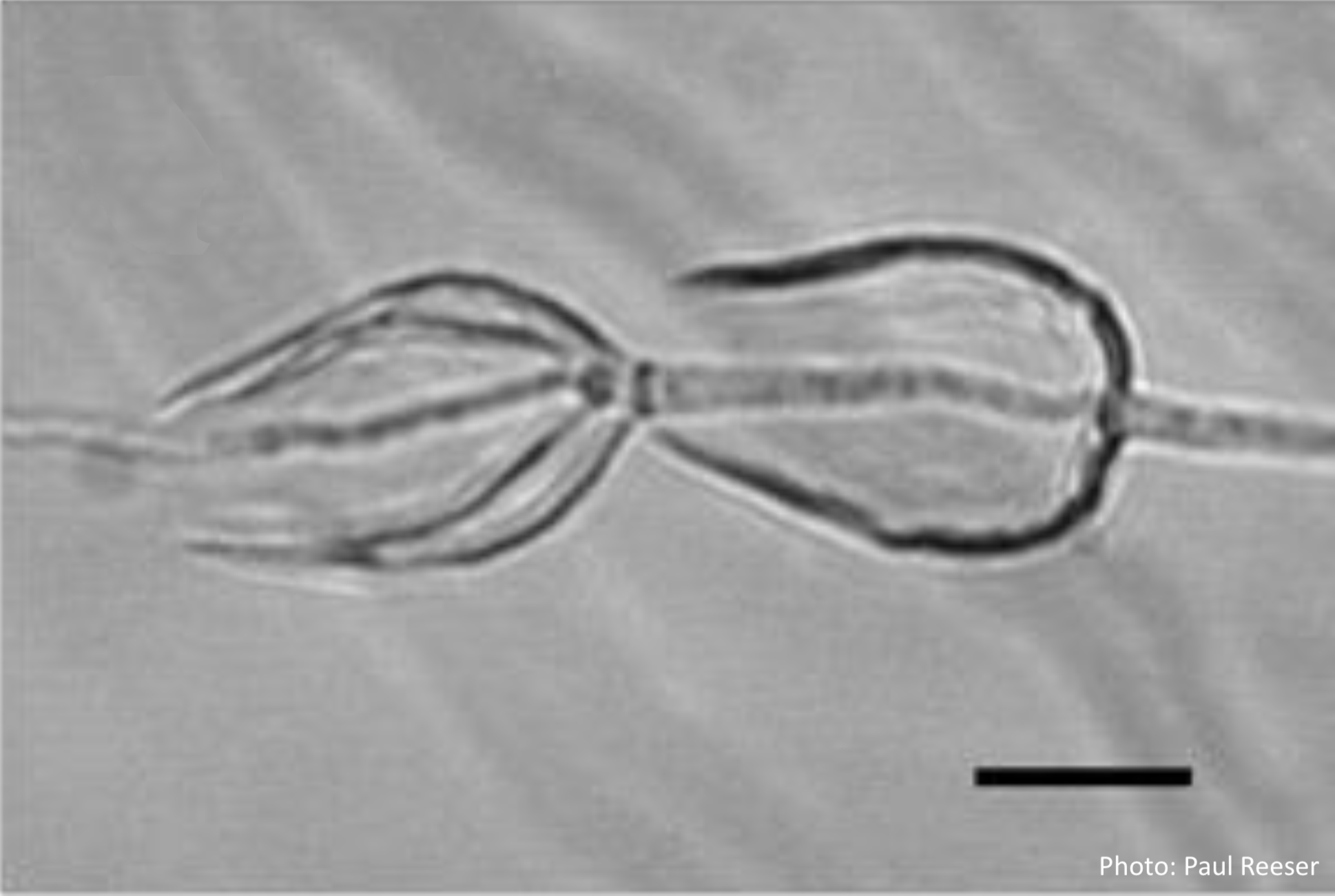 Phytophthora chlamydospora sporangia in water, showing internal proliferation. Bar is 20 µm. |
P. tentaculata hyphae 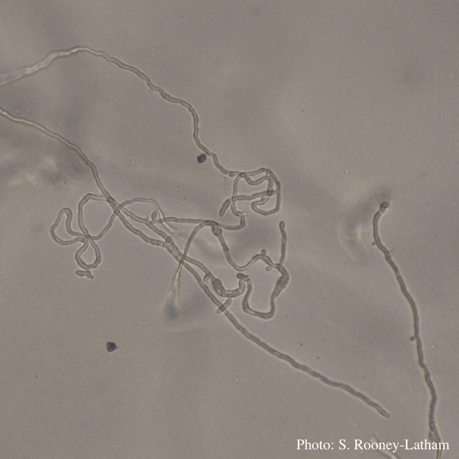 Looping hyphae commonly seen with P. tentaculata on PARP media |
P. ramorum chlamydospores 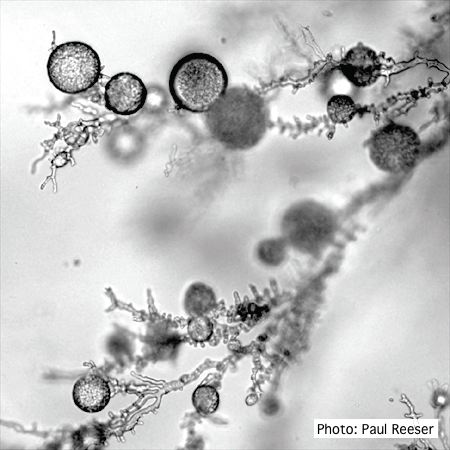 P. ramorum chlamydospores |