


Rosaceous colony morphology at 14 days at 20°C on PDA
Photo Gallery
Site will be retired 9/1/2026
This site is no longer being developed and will be retired on September 1, 2026. Please contact us if you have any questions or would like to provide support to continue the project.
|
P. cambivora colony morphology on PDA 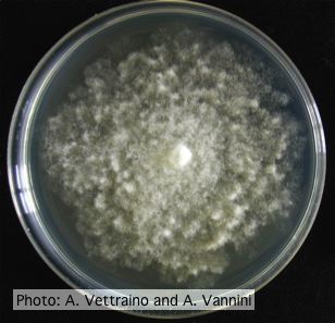 |
P. cactorum sporangia 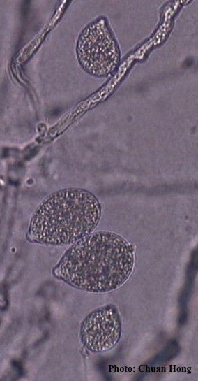 P. cactorum sporangia |
Comparative gametangial morphology of Phytophthora Clade 5 species 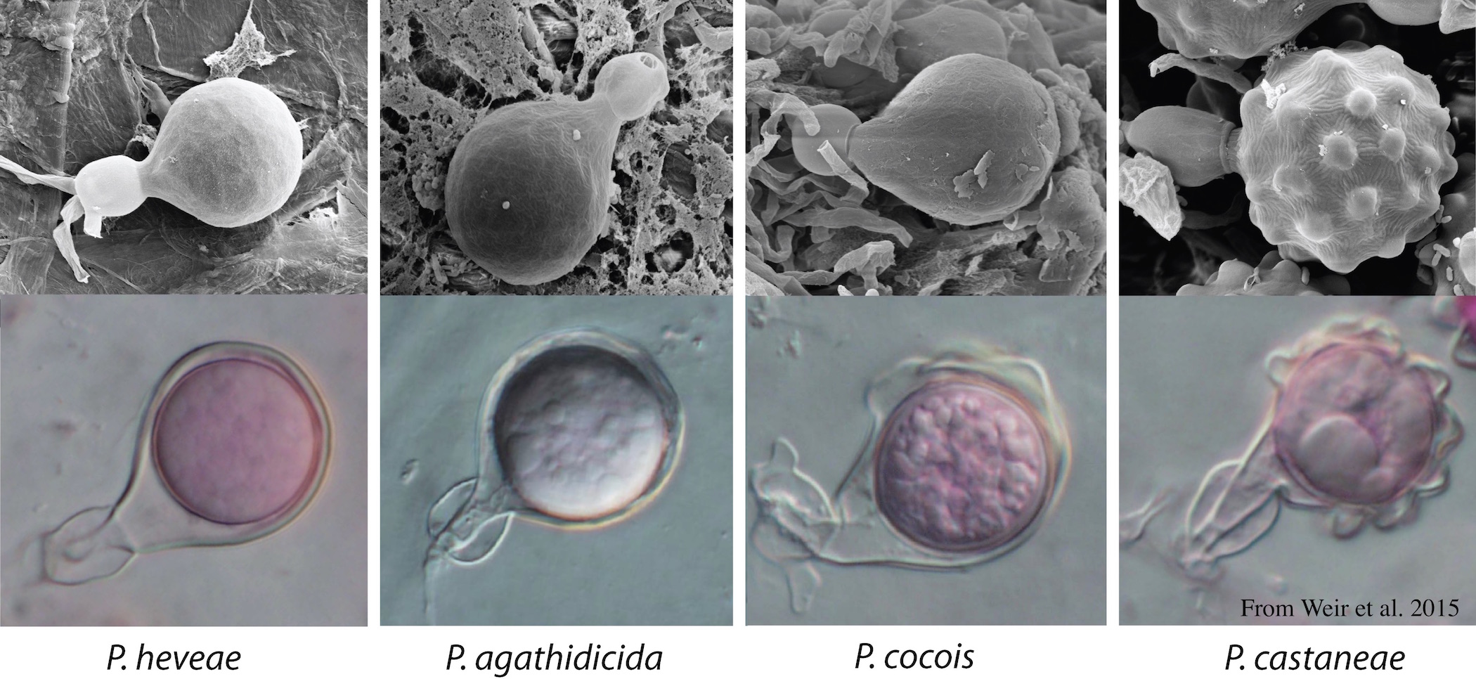 Comparative gametangial morphology of Phytophthora Clade 5 species, with SEM (top) and light microscopy (bottom). P. heveae has smooth walled oogonia with funnel-shaped, amphigynous antheridia. P. agathidicida has mildly stipulate oogonia with globose amphigynous antheridia. P.cocois has mildly bullate oogonia with reflexed amphigynous antheridia. P. castaneae has coarsely bullate oogonium with rugose protuberances and narrow amphigynous antheridia (Weir et al. 2015). |
|
Growth of P. arenaria on V8 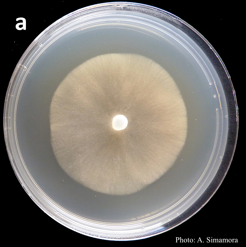 Colony morphology of Phytophthora arenaria after 7 days at 20°C on V8 agar |
P. pluvialis symptoms 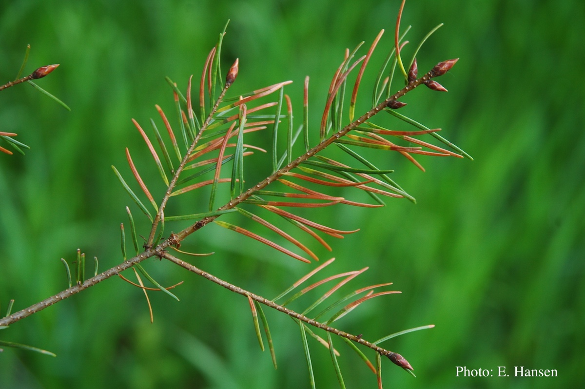 Symptoms of red needle cast on Douglas-fir needles |
P. ramorum sporangium 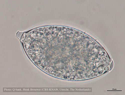 Deciduous sporangium, photo from Q-bank, used with permission |
|
Boot wash to station to control spread of P. agathidicida 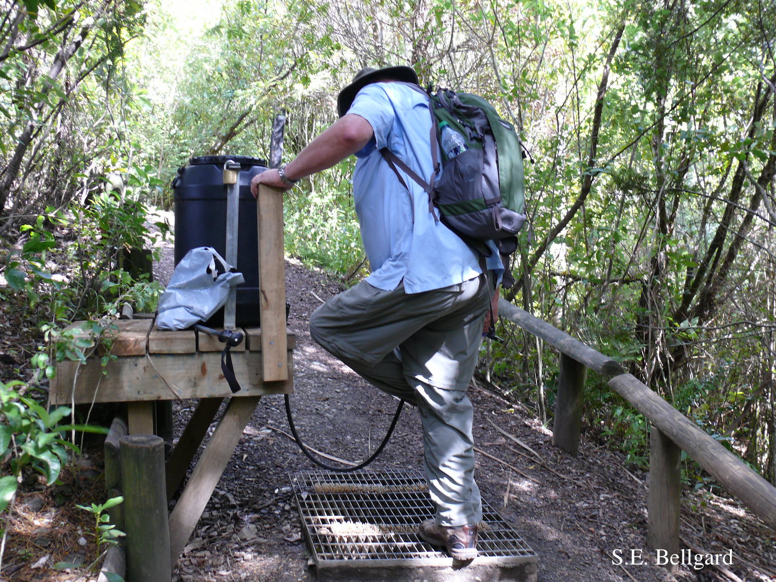 Use of hypochlorite solution applied through a “livestock drench-gun”, integrated with a soil grate to allow potentially contaminated soil to be collected |
P. nemorosa colony morphology on V8 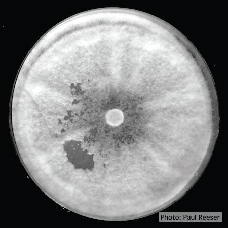 Colony morphology on V8 at 14 days |
P. tentaculata disease symptoms on sticky monkey flower 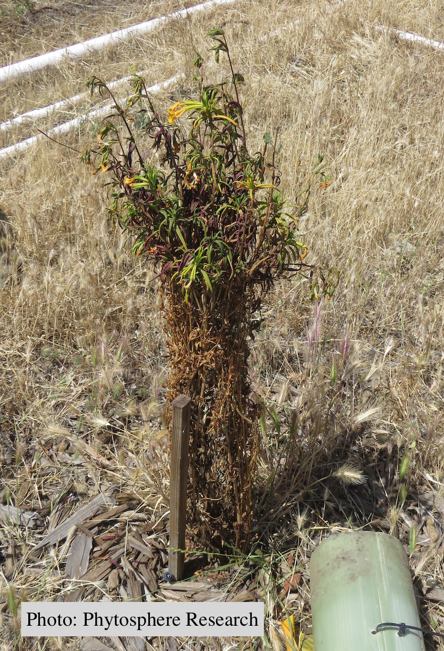 Outplanted sticky monkey flower (Diplacus aurantiacus) infected with P. |
|
P. cambivora sporangia 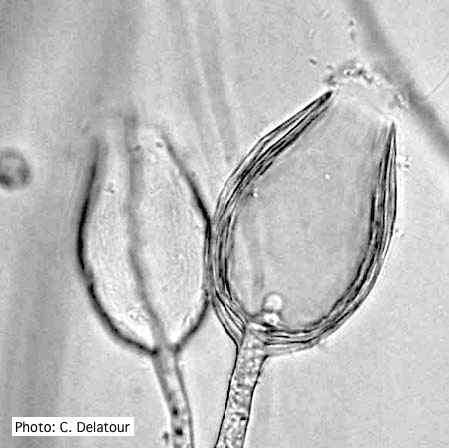 Empty sporangia of P. cambivora showing nested internal proliferation |
P. cactorum bleeding canker 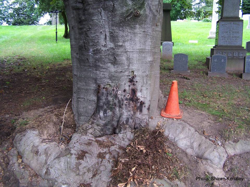 Bleeding canker on European beech (Fagus sylvatica) |
P. austrocedrae - hyphal swellings 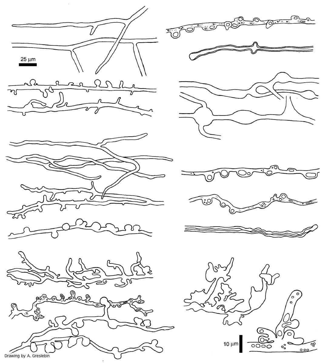 Morphology of hyphae of Phytophthora austrocedrae, from Greslebin et al. 2007 |