
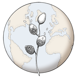

Oogonium with amphigynous antheridia, photo from Q-bank, used with permission
Photo Gallery
Site will be retired 9/1/2026
This site is no longer being developed and will be retired on September 1, 2026. Please contact us if you have any questions or would like to provide support to continue the project.
|
P. kernoviae oogonia 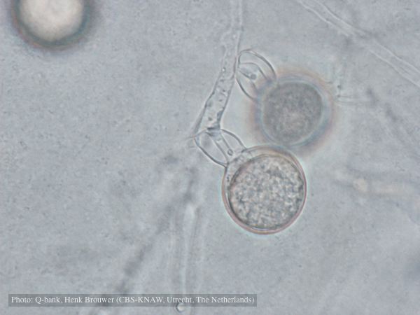 |
P. kernoviae sporangium 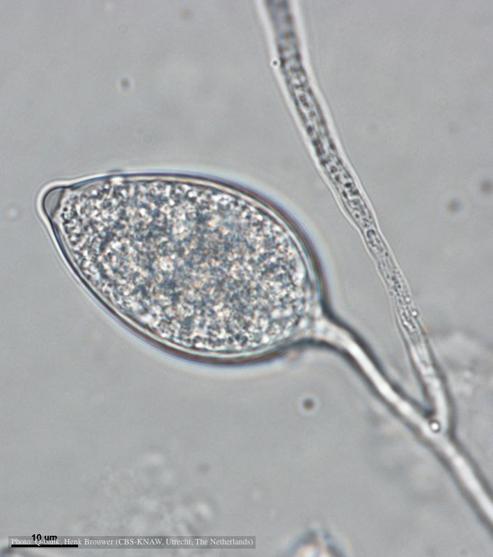 Papillate and caducous sporangium, photo from Q-bank, used with permission |
P. kernoviae colony morphology 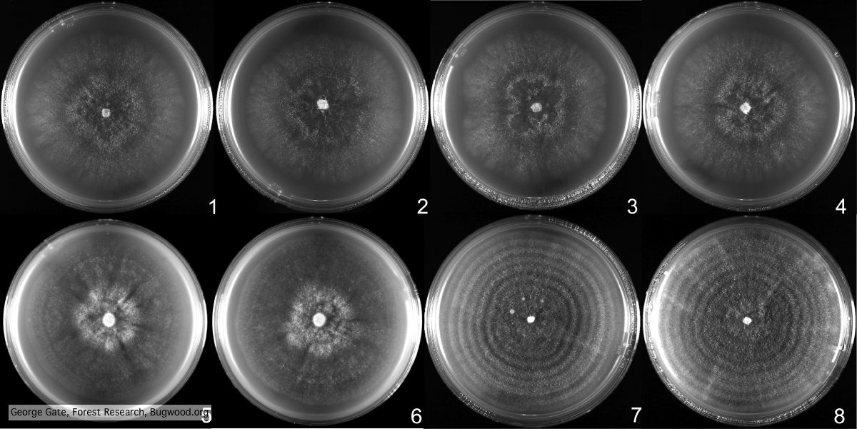 From Mycol.Res 109, 853-859; growth on CA under different conditions |
|
P. kernoviae sporangia 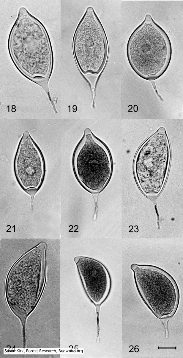 Mycol.Res 109, 853-859; Figs 18-22. Regular, ovoid limoniform sporangia. Figs 23-26. Asymmetrical or sporangia |
P. kernoviae canker 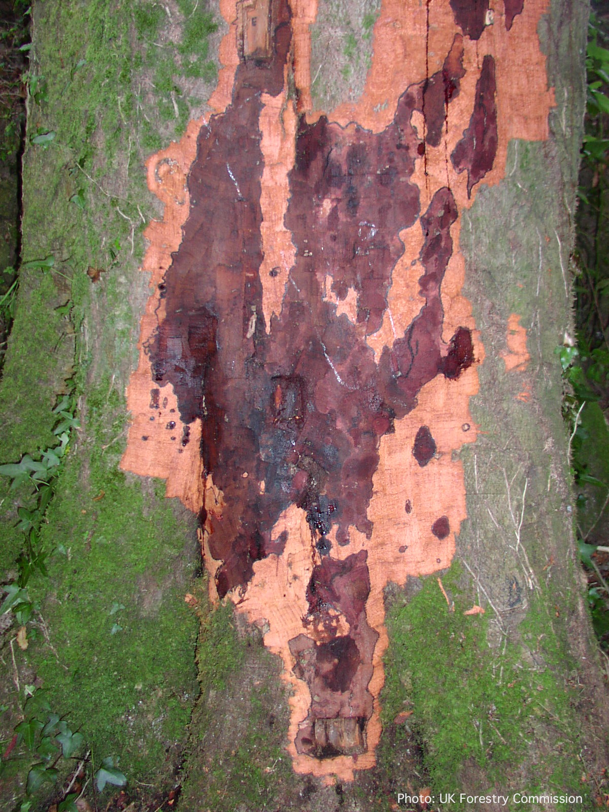 Bole lesion on Fagus sylvatica |
P. kernoviae disease on beech 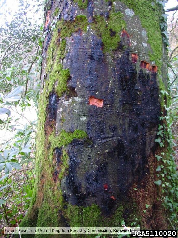 External lesion; 14 November 2003 |
|
P. kernoviae disease on European beech 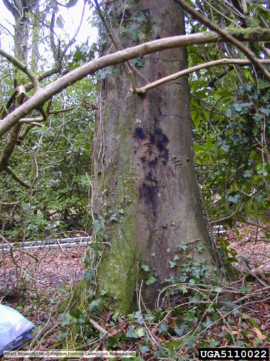 Bleeding lesion on trunk of Fagus sylvatica |
P. kernoviae leaf wilt 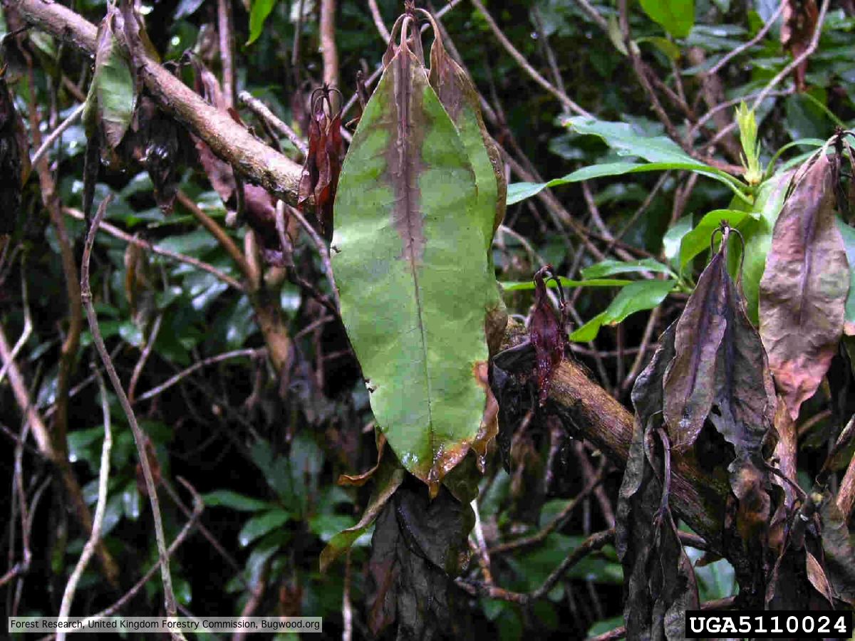 Wilted leaf of infected rhododendron |
P. kernoviae colony morphology on V8 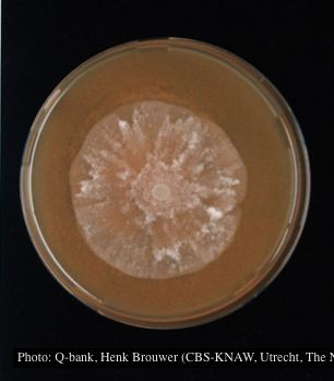 Colony morphology at 7 days at 18°C on V8, photo from Q-bank, used with permission. |
|
P. kernoviae leaf wilt 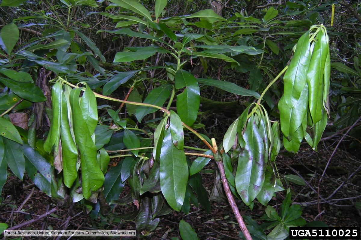 Wilted leaf of infected rhododendron |
P. kernoviae sporangium 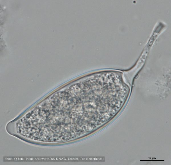 Asymmetrical sporangium, photo from Q-bank, used with permission |
P. kernoviae oogonia 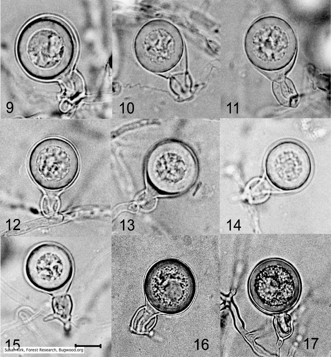 Mycol.Res 109, 853-859; Representative oogonia, antheridia and thick walled plerotic oospores of Phytophthora kernoviae. |