

Gum oozing out of longitudinal lesion
Photo Gallery
Site will be retired 9/1/2026
This site is no longer being developed and will be retired on September 1, 2026. Please contact us if you have any questions or would like to provide support to continue the project.
|
P. agathidicida lesion on kauri tree 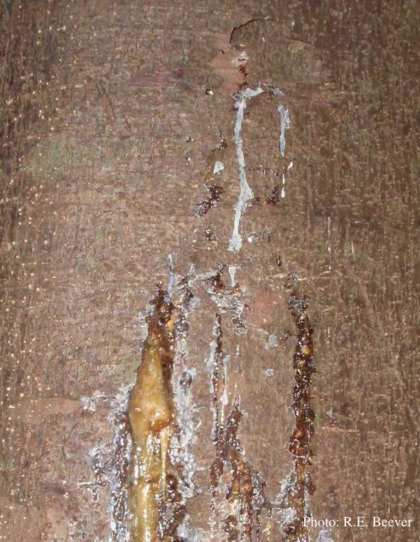 |
P. pseudosyringae oogonium 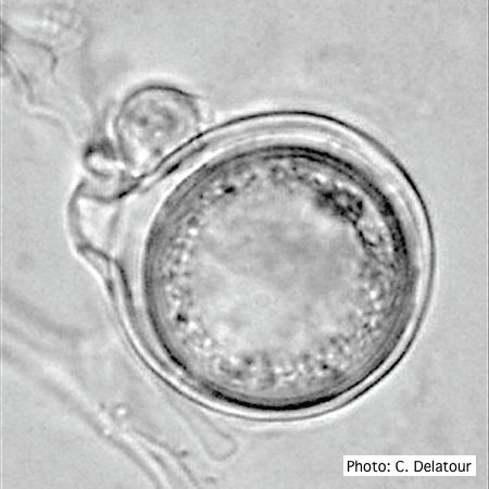 Oogonium with paragynous antheridium in agar |
P. cinnamomi on Banksia 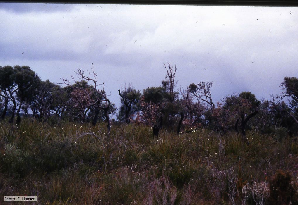 Death of woodland Banksia, Western Australia |
|
P. arenaria oogonium 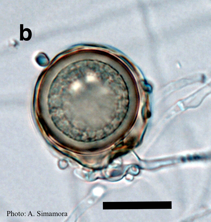 Aplerotic oogonia of P. arenaria with paragynous antheridia. Scale bar = 20 μm |
P. palmivora sporangia, chlamydospores, hyphae 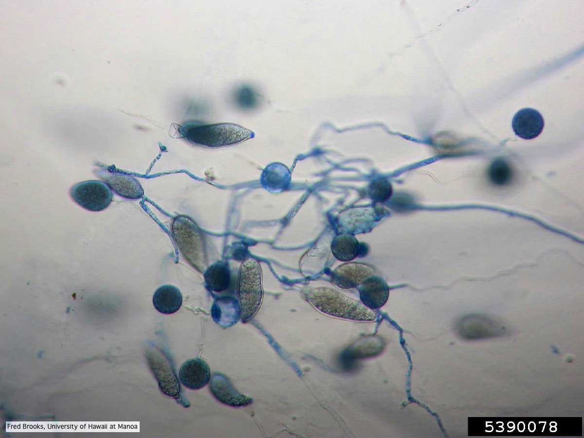 Sporangia (sporangiospores), chlamydospores, and hyphae stained with Cotton Blue |
P. pinifolia hyphal growth 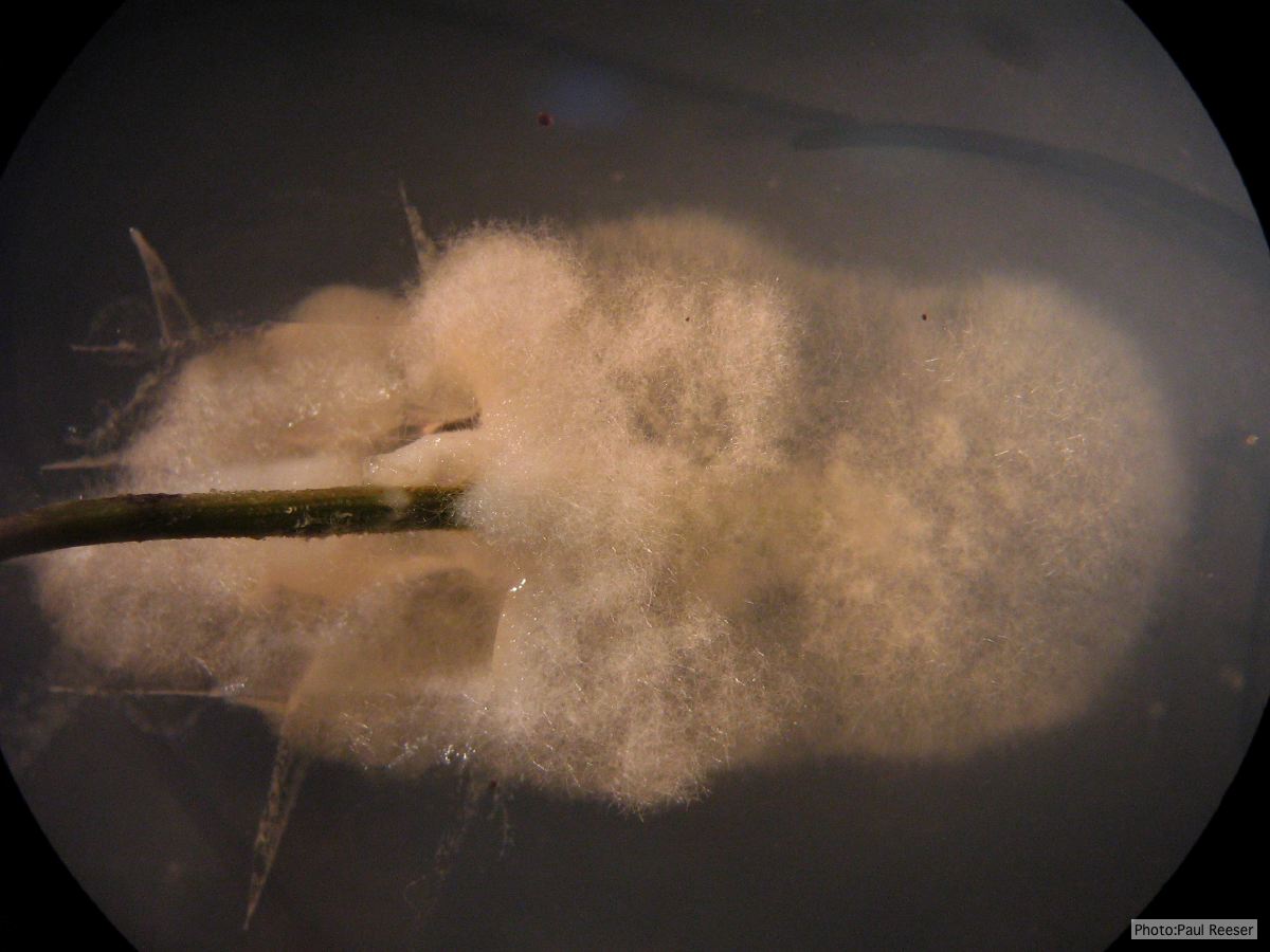 P. pinifolia pathogen growing from infected needle on selective agar |
|
P. kernoviae disease on beech 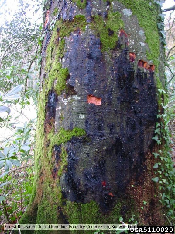 External lesion; 14 November 2003 |
P. cambivora colony morphology on PDA 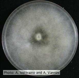 Uniform fluffy colony morphology at 14 days at 20°C on PDA |
P. chlamydospora sporangium 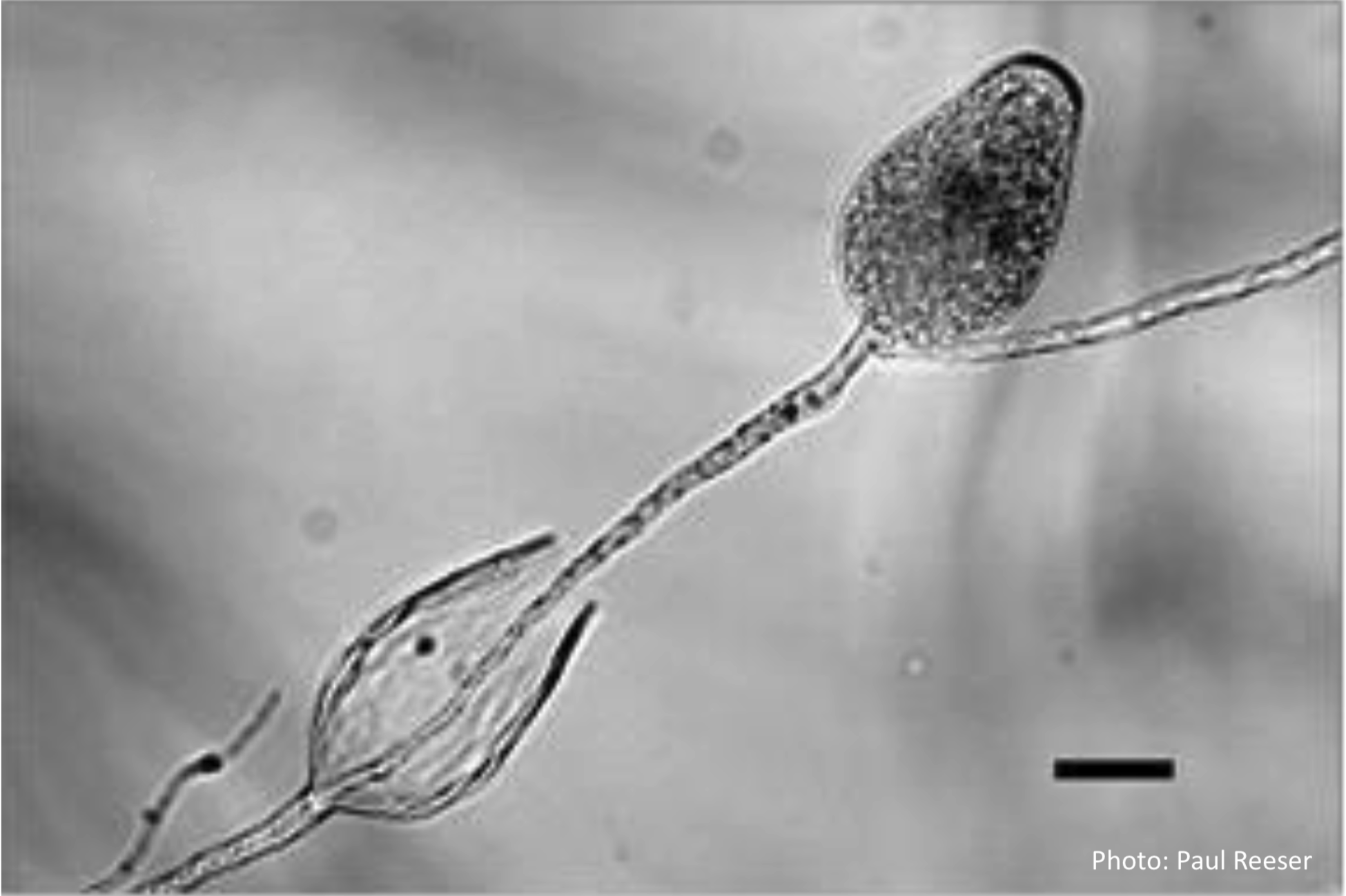 Phytophthora chlamydospora sporangia in water, showing subsporangial elongation. Bar is 20 µm. |
|
Necrotic lesion in phloem caused by P. austrocedrae 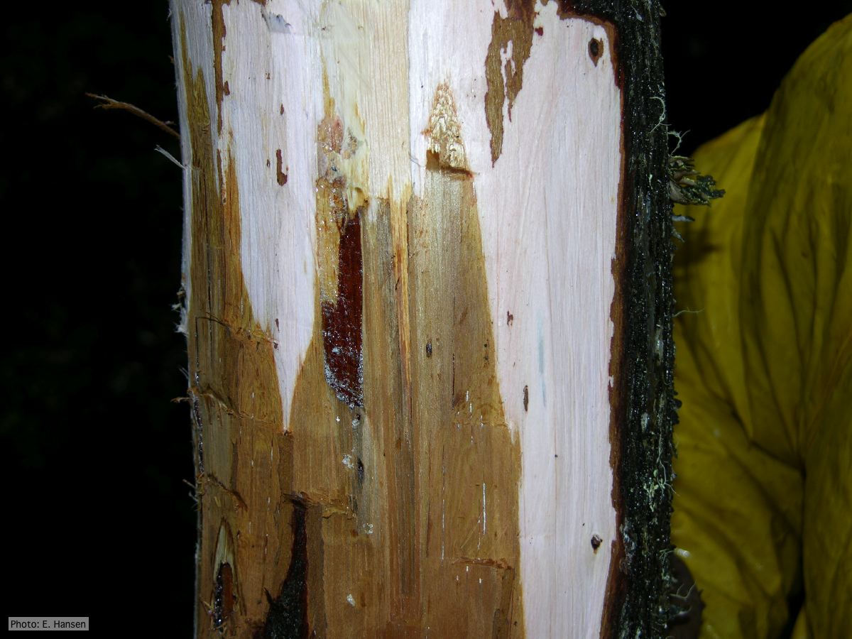 Necrotic lesion in phloem with resin pocket caused by P. austrocedrae |
P. alni symptoms on European Alder 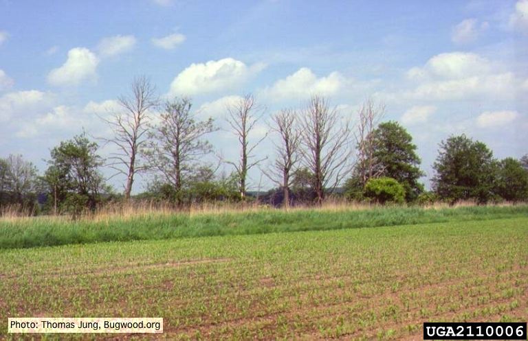 Mature, riparian common alder (A. glutinosa) stand with high impact of Phytophthora root and collar rot. |
P. lateralis sporangia 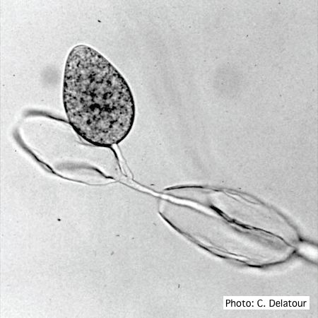 Sporangia of P. lateralis showing internal and external proliferation |