


Colony morphology of P. pinifolia at 20°C on CMA-NARP after 3 weeks. From Duran et al. 2008
Photo Gallery
Site will be retired 9/1/2026
This site is no longer being developed and will be retired on September 1, 2026. Please contact us if you have any questions or would like to provide support to continue the project.
|
P. pinifolia colony morphology on CMA-NARP 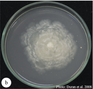 |
P. pinifolia on Pinus radiata 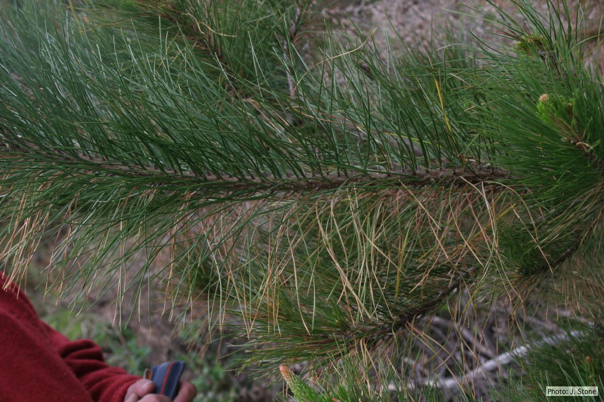 Dead needles on lower side of P. radiata branch. |
P. pinifolia on Pinus radiata 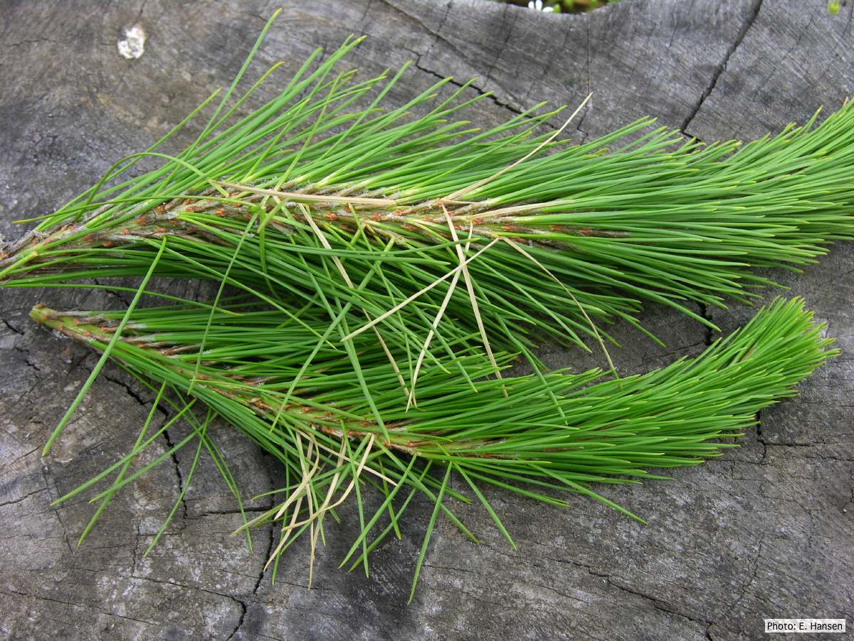 Pinus radiata, note Infected needles at right angles to stem |
|
P. pinifolia colony morphology on V8 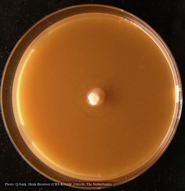 Colony pattern after 7 days on V8 at 24C, photo from Q-bank, used with permission |
P. pinifolia sporangia 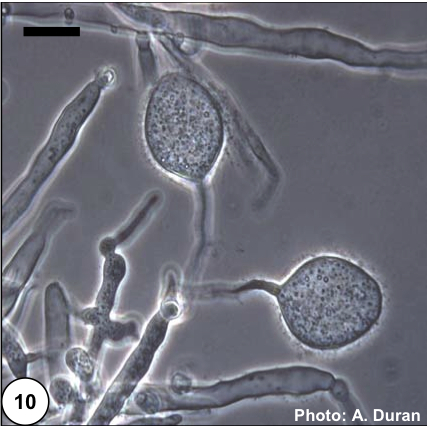 Non- papillate and caducous sporangia of Phytophthora pinifolia isolated from the infected P. radiata needles. |
P. pinifolia hyphal growth 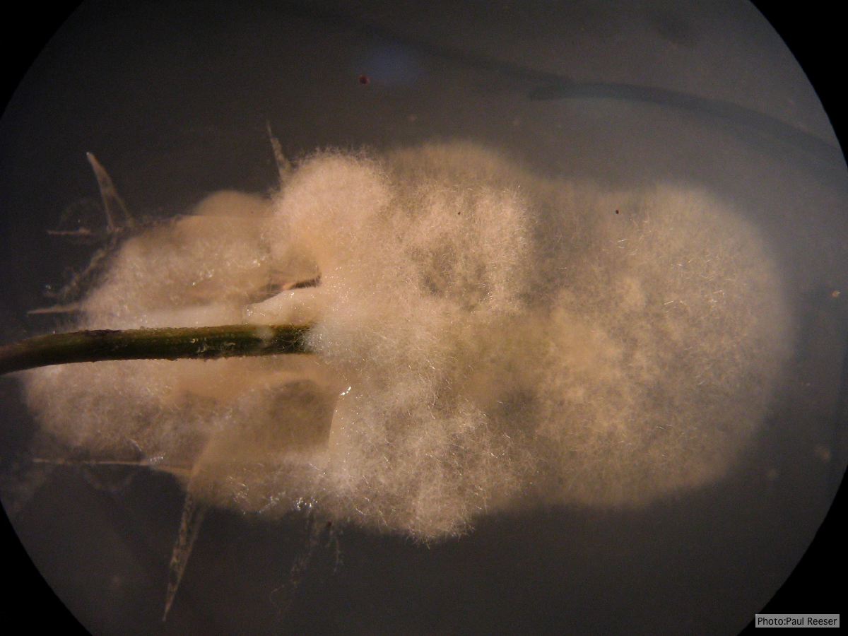 P. pinifolia pathogen growing from infected needle on selective agar |
|
P. pinifolia hyphal swellings 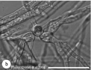 Spherical hyphal swelling with radiating hyphae (from Duran et al. 2008). Scale bar = 20 μm. |
P. pinifolia on Pinus radiata 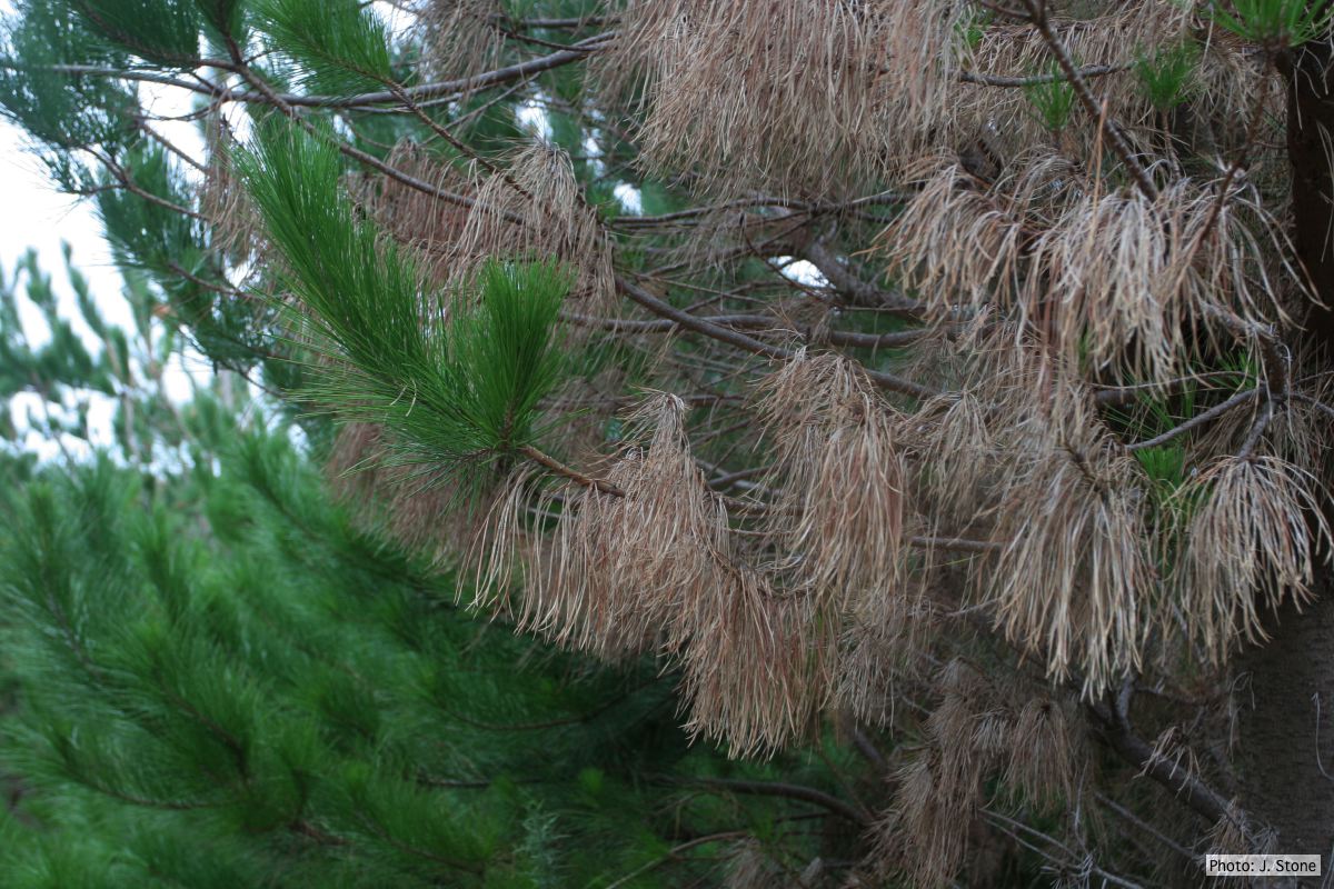 Pinus radiata dead needles caused by DFP with healthy new growth from DFP |
P. pinifolia colony morphology on PDA 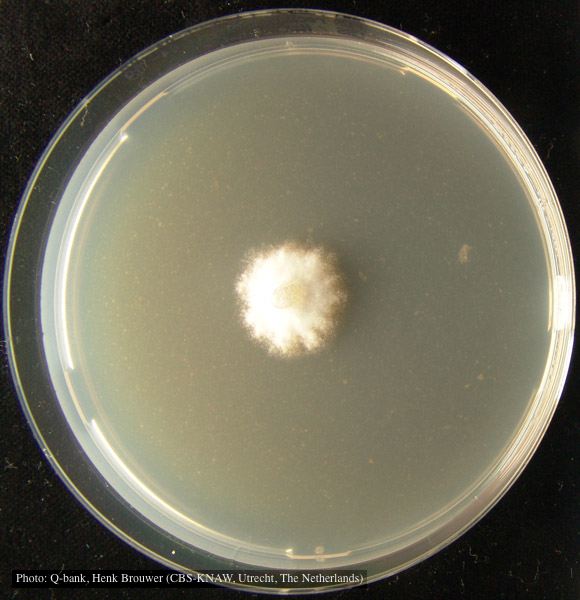 Colony pattern after 7 days on PDA at 24C, photo from Q-bank, used with permission. |
|
P. pinifolia on Pinus radiata 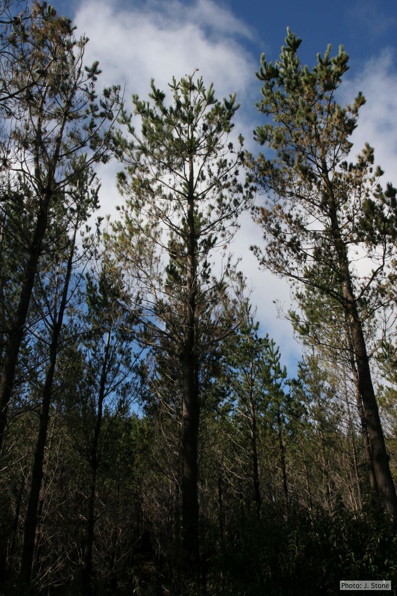 Pinus radiata impacted by DFP, note healthy new growth |
P. pinifolia on Pinus radiata 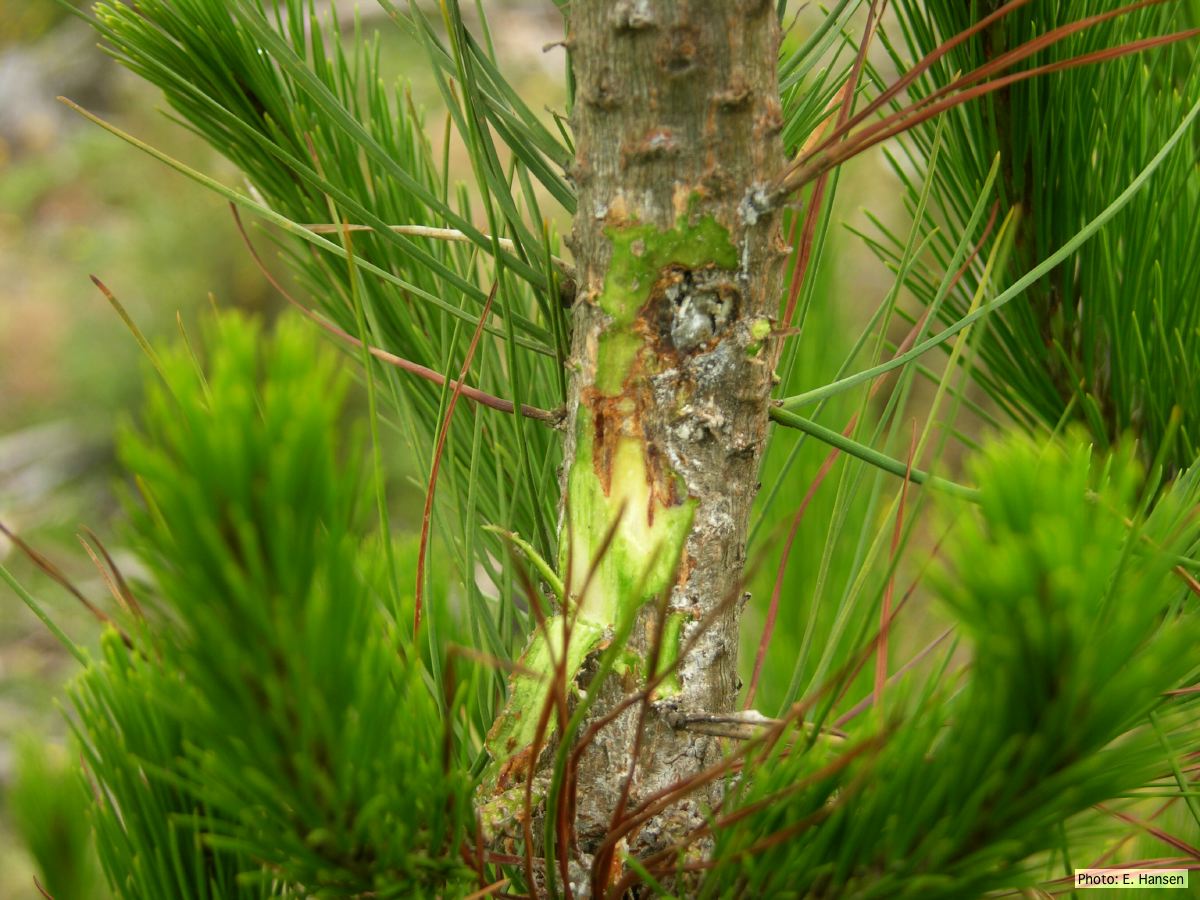 Pinus radiata, note Stem canker associated with necrotic needles. |
P. pinifolia sporangium 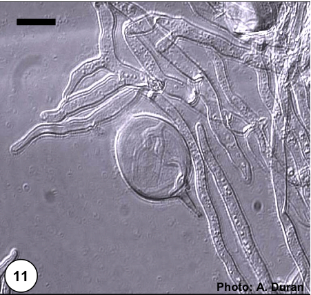 Non- papillate and caducous sporangia of Phytophthora pinifolia isolated from the infected P. radiata needles. |